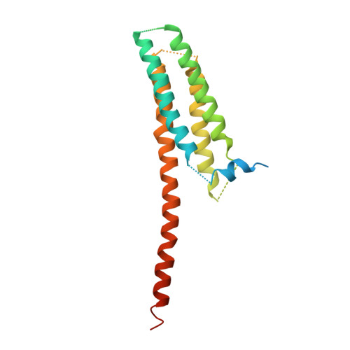
3WKV | pdb_00003wkv

Voltage-gated proton channel: VSOP/Hv1 chimeric channel


Experimental Data Snapshot

  • Method: X-RAY DIFFRACTION
  • Resolution: 3.45 Å
  • R-Value Free: 
    0.357 (Depositor), 0.362 (DCC) 
  • R-Value Work: 
    0.341 (Depositor), 0.341 (DCC) 
  • R-Value Observed: 
    0.342 (Depositor) 

wwPDB Validation 3D Report Full Report

Validation slider image for 3WKV

This is version 1.2 of the entry. See complete history. 

Literature

X-ray crystal structure of voltage-gated proton channel.

Takeshita, K., Sakata, S., Yamashita, E., Fujiwara, Y., Kawanabe, A., Kurokawa, T., Okochi, Y., Matsuda, M., Narita, H., Okamura, Y., Nakagawa, A.

(2014) Nat Struct Mol Biol 21: 352-357

  • DOI: https://doi.org/10.1038/nsmb.2783
  • Primary Citation Related Structures: 
    3WKV

  • PubMed Abstract: 

    The voltage-gated proton channel Hv1 (or VSOP) has a voltage-sensor domain (VSD) with dual roles of voltage sensing and proton permeation. Its gating is sensitive to pH and Zn(2+). Here we present a crystal structure of mouse Hv1 in the resting state at 3.45-Å resolution. The structure showed a 'closed umbrella' shape with a long helix consisting of the cytoplasmic coiled coil and the voltage-sensing helix, S4, and featured a wide inner-accessible vestibule. Two out of three arginines in S4 were located below the phenylalanine constituting the gating charge-transfer center. The extracellular region of each protomer coordinated a Zn(2+), thus suggesting that Zn(2+) stabilizes the resting state of Hv1 by competing for acidic residues that otherwise form salt bridges with voltage-sensing positive charges on S4. These findings provide a platform for understanding the general principles of voltage sensing and proton permeation.


  • Organizational Affiliation: 
    • 1] Institute for Protein Research, Osaka University, Suita, Japan. [2] Graduate School of Medicine, Osaka University, Suita, Japan. [3] Institute for Academic Initiatives, Osaka University, Suita, Japan.

Macromolecule Content 

  • Total Structure Weight: 22.88 kDa 
  • Atom Count: 1,085 
  • Modeled Residue Count: 140 
  • Deposited Residue Count: 196 
  • Unique protein chains: 1

Macromolecules

Find similar proteins by:|  3D Structure
Entity ID: 1
MoleculeChains  Sequence LengthOrganismDetailsImage
Ion channel196Mus musculusMutation(s): 0 
Membrane Entity: Yes 
UniProt
Find proteins for Q3U2S8 (Mus musculus)
Explore Q3U2S8 
Go to UniProtKB:  Q3U2S8
Entity Groups
Sequence Clusters30% Identity50% Identity70% Identity90% Identity95% Identity100% Identity
UniProt GroupQ3U2S8
Sequence Annotations
Expand
Reference Sequence

Experimental Data & Validation

Experimental Data

  • Method: X-RAY DIFFRACTION
  • Resolution: 3.45 Å
  • R-Value Free:  0.357 (Depositor), 0.362 (DCC) 
  • R-Value Work:  0.341 (Depositor), 0.341 (DCC) 
  • R-Value Observed: 0.342 (Depositor) 
Space Group: P 63
Unit Cell:
Length ( Å )Angle ( ˚ )
a = 86.54α = 90
b = 86.54β = 90
c = 89.553γ = 120
Software Package:
Software NamePurpose
DENZOdata reduction
SCALEPACKdata scaling
SHELXphasing
PHENIXrefinement
PDB_EXTRACTdata extraction
BSSdata collection
SHELXDphasing

Structure Validation

View Full Validation Report


Entry History 

Deposition Data

Revision History  (Full details and data files)

  • Version 1.0: 2014-03-05
    Type: Initial release
  • Version 1.1: 2020-01-01
    Changes: Database references
  • Version 1.2: 2024-03-20
    Changes: Data collection, Database references