3WIA | pdb_00003wia

Crystal structure of the N-terminal 1-37 residues deleted mutant of Geobacillus copper nitrite reductase


Experimental Data Snapshot

  • Method: X-RAY DIFFRACTION
  • Resolution: 1.77 Å
  • R-Value Free: 
    0.192 (Depositor), 0.191 (DCC) 
  • R-Value Work: 
    0.171 (Depositor), 0.171 (DCC) 
  • R-Value Observed: 
    0.172 (Depositor) 

Starting Model: experimental
View more details

wwPDB Validation 3D Report Full Report

Validation slider image for 3WIA

This is version 1.1 of the entry. See complete history

Literature

Structural and functional characterization of the Geobacillus copper nitrite reductase: involvement of the unique N-terminal region in the interprotein electron transfer with its redox partner

Fukuda, Y.Koteishi, H.Yoneda, R.Tamada, T.Takami, H.Inoue, T.Nojiri, M.

(2014) Biochim Biophys Acta 1837: 396-405

  • DOI: https://doi.org/10.1016/j.bbabio.2014.01.004
  • Primary Citation Related Structures: 
    3WI9, 3WIA

  • PubMed Abstract: 

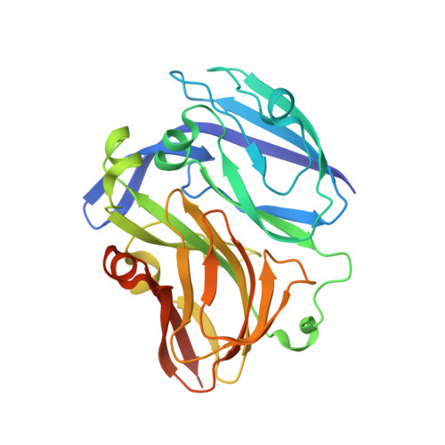
    The crystal structures of copper-containing nitrite reductase (CuNiR) from the thermophilic Gram-positive bacterium Geobacillus kaustophilus HTA426 and the amino (N)-terminal 68 residue-deleted mutant were determined at resolutions of 1.3Å and 1.8Å, respectively. Both structures show a striking resemblance with the overall structure of the well-known CuNiRs composed of two Greek key β-barrel domains; however, a remarkable structural difference was found in the N-terminal region. The unique region has one β-strand and one α-helix extended to the northern surface of the type-1 copper site. The superposition of the Geobacillus CuNiR model on the electron-transfer complex structure of CuNiR with the redox partner cytochrome c551 in other denitrifier system led us to infer that this region contributes to the transient binding with the partner protein during the interprotein electron transfer reaction in the Geobacillus system. Furthermore, electron-transfer kinetics experiments using N-terminal residue-deleted mutant and the redox partner, Geobacillus cytochrome c551, were carried out. These structural and kinetics studies demonstrate that the region is directly involved in the specific partner recognition.


  • Organizational Affiliation
    • Department of Chemistry, Graduate School of Science, Osaka University, 1-1 Machikaneyama, Toyonaka, Osaka 560-0043, Japan; Department of Materials Chemistry, Graduate School of Engineering, Osaka University, Suita, Osaka 565-0871, Japan; Molecular Biology Research Center, Quantum Beam Science Directorate, Japan Atomic Energy Agency, Tokai, Ibaraki 319-1195, Japan.

Macromolecule Content 

  • Total Structure Weight: 287.2 kDa 
  • Atom Count: 21,279 
  • Modeled Residue Count: 2,456 
  • Deposited Residue Count: 2,583 
  • Unique protein chains: 1

Macromolecules

Find similar proteins by:|  3D Structure
Entity ID: 1
MoleculeChains  Sequence LengthOrganismDetailsImage
Nitrite reductase
A, B, C, D, E
A, B, C, D, E, F, G, H, I
287Geobacillus kaustophilus HTA426Mutation(s): 0 
Gene Names: GK0767
EC: 1.7.2.1
UniProt
Find proteins for Q5L1X8 (Geobacillus kaustophilus (strain HTA426))
Explore Q5L1X8 
Go to UniProtKB:  Q5L1X8
Entity Groups
Sequence Clusters30% Identity50% Identity70% Identity90% Identity95% Identity100% Identity
UniProt GroupQ5L1X8
Sequence Annotations
Expand
Reference Sequence

Small Molecules

Ligands 2 Unique
IDChains Name / Formula / InChI Key2D Diagram3D Interactions
CU

Query on CU


Download:Ideal Coordinates CCD File
AA [auth E]
BA [auth E]
EA [auth F]
FA [auth F]
HA [auth G]
AA [auth E],
BA [auth E],
EA [auth F],
FA [auth F],
HA [auth G],
IA [auth G],
J [auth A],
K [auth A],
MA [auth H],
NA [auth H],
O [auth B],
P [auth B],
QA [auth I],
RA [auth I],
S [auth C],
T [auth C],
V [auth D],
W [auth D]
COPPER (II) ION
Cu
JPVYNHNXODAKFH-UHFFFAOYSA-N
FMT

Query on FMT


Download:Ideal Coordinates CCD File
CA [auth E]
DA [auth E]
GA [auth F]
JA [auth G]
KA [auth G]
CA [auth E],
DA [auth E],
GA [auth F],
JA [auth G],
KA [auth G],
L [auth A],
LA [auth H],
M [auth A],
N [auth B],
OA [auth H],
PA [auth I],
Q [auth B],
R [auth C],
SA [auth I],
U [auth C],
X [auth D],
Y [auth D],
Z [auth D]
FORMIC ACID
C H2 O2
BDAGIHXWWSANSR-UHFFFAOYSA-N

Experimental Data & Validation

Experimental Data

  • Method: X-RAY DIFFRACTION
  • Resolution: 1.77 Å
  • R-Value Free:  0.192 (Depositor), 0.191 (DCC) 
  • R-Value Work:  0.171 (Depositor), 0.171 (DCC) 
  • R-Value Observed: 0.172 (Depositor) 
Space Group: P 1 21 1
Unit Cell:
Length ( Å )Angle ( ˚ )
a = 89.317α = 90
b = 164.991β = 102.27
c = 126.561γ = 90
Software Package:
Software NamePurpose
HKL-2000data collection
MOLREPphasing
REFMACrefinement
HKL-2000data reduction
SCALEPACKdata scaling

Structure Validation

View Full Validation Report


Entry History 

Deposition Data

Revision History  (Full details and data files)

  • Version 1.0: 2014-07-23
    Type: Initial release
  • Version 1.1: 2023-11-08
    Changes: Data collection, Database references, Derived calculations, Refinement description