3WG5 | pdb_00003wg5

1510-N membrane-bound stomatin-specific protease K138A mutant in complex with a substrate peptide under heat treatment


Experimental Data Snapshot

  • Method: X-RAY DIFFRACTION
  • Resolution: 2.40 Å
  • R-Value Free: 
    0.241 (Depositor), 0.241 (DCC) 
  • R-Value Work: 
    0.202 (Depositor), 0.202 (DCC) 
  • R-Value Observed: 
    0.206 (Depositor) 

Starting Model: experimental
View more details

wwPDB Validation 3D Report Full Report

Validation slider image for 3WG5

This is version 1.2 of the entry. See complete history

Literature

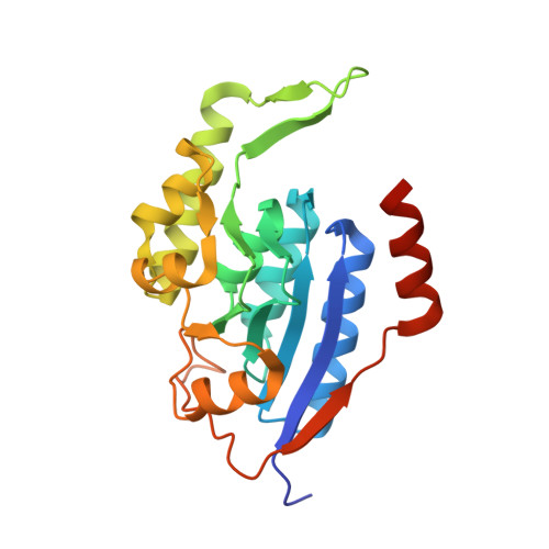
Structural and biochemical analysis of a thermostable membrane-bound stomatin-specific protease.

Yokoyama, H.Kobayashi, D.Takizawa, N.Fujii, S.Matsui, I.

(2013) J Synchrotron Radiat 20: 933-937

  • DOI: https://doi.org/10.1107/S0909049513021328
  • Primary Citation Related Structures: 
    3WG5

  • PubMed Abstract: 

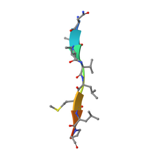
    Membrane-bound proteases are involved in various regulatory functions. The N-terminal region of PH1510p (1510-N) from the hyperthermophilic archaeon Pyrococcus horikoshii is a serine protease with a catalytic Ser-Lys dyad (Ser97 and Lys138), and specifically cleaves the C-terminal hydrophobic region of the p-stomatin PH1511p. In a form of human hemolytic anemia known as hereditary stomatocytosis, the stomatin protein is deficient in the erythrocyte membrane due to mis-trafficking. In order to understand the catalytic mechanism of 1510-N in more detail, here the structural and biochemical analysis of 1510-N is reported. Two degraded products were produced via acyl-enzyme intermediates. 1510-N is a thermostable protease, and thus crystallization after heat treatment of the protease-peptide complex was attempted in order to understand the catalytic mechanism of 1510-N. The structure after heat treatment is almost identical to that with no heat treatment. According to the superposition between the structures with heat treatment and with no heat treatment, the N-terminal half of the peptide is superposed well, whereas the C-terminal half of the peptide is slightly deviated. The N-terminal half of the peptide binds to 1510-N more tightly than the C-terminal half of the peptide. The flexible L2 loops of 1510-N cover the peptide, and are involved in the protease activity.


  • Organizational Affiliation
    • School of Pharmaceutical Sciences, University of Shizuoka, 52-1 Yada, Suruga-ku, Shizuoka 422-8526, Japan.

Macromolecule Content 

  • Total Structure Weight: 52.32 kDa 
  • Atom Count: 3,603 
  • Modeled Residue Count: 446 
  • Deposited Residue Count: 470 
  • Unique protein chains: 2

Macromolecules

Find similar proteins by:|  3D Structure
Entity ID: 1
MoleculeChains  Sequence LengthOrganismDetailsImage
441aa long hypothetical nfeD protein
A, B
230Pyrococcus horikoshii OT3Mutation(s): 1 
Gene Names: PH1510
EC: 3.4.21
UniProt
Find proteins for O59179 (Pyrococcus horikoshii (strain ATCC 700860 / DSM 12428 / JCM 9974 / NBRC 100139 / OT-3))
Explore O59179 
Go to UniProtKB:  O59179
Entity Groups
Sequence Clusters30% Identity50% Identity70% Identity90% Identity95% Identity100% Identity
UniProt GroupO59179
Sequence Annotations
Expand
Reference Sequence
Find similar proteins by:  Sequence   |   3D Structure  
Entity ID: 2
MoleculeChains  Sequence LengthOrganismDetailsImage
PH1511 stomatin10Pyrococcus horikoshii OT3Mutation(s): 0 
UniProt
Find proteins for O59180 (Pyrococcus horikoshii (strain ATCC 700860 / DSM 12428 / JCM 9974 / NBRC 100139 / OT-3))
Explore O59180 
Go to UniProtKB:  O59180
Entity Groups
Sequence Clusters30% Identity50% Identity70% Identity90% Identity95% Identity100% Identity
UniProt GroupO59180
Sequence Annotations
Expand
Reference Sequence

Experimental Data & Validation

Experimental Data

  • Method: X-RAY DIFFRACTION
  • Resolution: 2.40 Å
  • R-Value Free:  0.241 (Depositor), 0.241 (DCC) 
  • R-Value Work:  0.202 (Depositor), 0.202 (DCC) 
  • R-Value Observed: 0.206 (Depositor) 
Space Group: P 43 21 2
Unit Cell:
Length ( Å )Angle ( ˚ )
a = 111.448α = 90
b = 111.448β = 90
c = 91.738γ = 90
Software Package:
Software NamePurpose
HKL-2000data collection
MOLREPphasing
REFMACrefinement
HKL-2000data reduction
HKL-2000data scaling

Structure Validation

View Full Validation Report


Entry History 

Deposition Data

Revision History  (Full details and data files)

  • Version 1.0: 2013-10-23
    Type: Initial release
  • Version 1.1: 2020-01-01
    Changes: Database references
  • Version 1.2: 2023-11-08
    Changes: Data collection, Database references, Derived calculations, Refinement description