3WFX | pdb_00003wfx

Crystal Structure of the Imidazole-Bound Form of the HGbRL's Globin Domain


Experimental Data Snapshot

  • Method: X-RAY DIFFRACTION
  • Resolution: 1.94 Å
  • R-Value Free: 
    0.234 (Depositor), 0.235 (DCC) 
  • R-Value Work: 
    0.183 (Depositor), 0.184 (DCC) 
  • R-Value Observed: 
    0.186 (Depositor) 

Starting Model: experimental
View more details

wwPDB Validation 3D Report Full Report

Validation slider image for 3WFX

Ligand Structure Quality Assessment 


This is version 1.2 of the entry. See complete history

Literature

Open and Lys-His Hexacoordinated Closed Structures of a Globin with Swapped Proximal and Distal Sites.

Teh, A.H.Saito, J.A.Najimudin, N.Alam, M.

(2015) Sci Rep 5: 11407-11407

  • DOI: https://doi.org/10.1038/srep11407
  • Primary Citation Related Structures: 
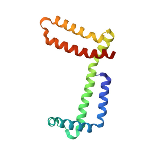
    3WFW, 3WFX

  • PubMed Abstract: 

    Globins are haem-binding proteins with a conserved fold made up of α-helices and can possess diverse properties. A putative globin-coupled sensor from Methylacidiphilum infernorum, HGbRL, contains an N-terminal globin domain whose open and closed structures reveal an untypical dimeric architecture. Helices E and F fuse into an elongated helix, resulting in a novel site-swapped globin fold made up of helices A-E, hence the distal site, from one subunit and helices F-H, the proximal site, from another. The open structure possesses a large cavity binding an imidazole molecule, while the closed structure forms a unique Lys-His hexacoordinated species, with the first turn of helix E unravelling to allow Lys52(E10) to bind to the haem. Ligand binding induces reorganization of loop CE, which is stabilized in the closed form, and helix E, triggering a large conformational movement in the open form. These provide a mechanical insight into how a signal may be relayed between the globin domain and the C-terminal domain of HGbRL, a Roadblock/LC7 domain. Comparison with HGbI, a closely related globin, further underlines the high degree of structural versatility that the globin fold is capable of, enabling it to perform a diversity of functions.


  • Organizational Affiliation
    • Centre for Chemical Biology, Universiti Sains Malaysia, 10 Persiaran Bukit Jambul, 11900 Bayan Lepas, Penang, Malaysia.

Macromolecule Content 

  • Total Structure Weight: 35.2 kDa 
  • Atom Count: 2,624 
  • Modeled Residue Count: 271 
  • Deposited Residue Count: 276 
  • Unique protein chains: 1

Macromolecules

Find similar proteins by:|  3D Structure
Entity ID: 1
MoleculeChains  Sequence LengthOrganismDetailsImage
Hemoglobin-like flavoprotein fused to Roadblock/LC7 domain
A, B
138Methylacidiphilum infernorum V4Mutation(s): 0 
Gene Names: hmpMinf_1089
UniProt
Find proteins for B3DUZ1 (Methylacidiphilum infernorum (isolate V4))
Explore B3DUZ1 
Go to UniProtKB:  B3DUZ1
Entity Groups
Sequence Clusters30% Identity50% Identity70% Identity90% Identity95% Identity100% Identity
UniProt GroupB3DUZ1
Sequence Annotations
Expand
Reference Sequence

Small Molecules

Ligands 3 Unique
IDChains Name / Formula / InChI Key2D Diagram3D Interactions
HEM

Query on HEM


Download:Ideal Coordinates CCD File
C [auth A],
I [auth B]
PROTOPORPHYRIN IX CONTAINING FE
C34 H32 Fe N4 O4
KABFMIBPWCXCRK-RGGAHWMASA-L
HEZ

Query on HEZ


Download:Ideal Coordinates CCD File
D [auth A]
E [auth A]
H [auth B]
J [auth B]
K [auth B]
D [auth A],
E [auth A],
H [auth B],
J [auth B],
K [auth B],
L [auth B]
HEXANE-1,6-DIOL
C6 H14 O2
XXMIOPMDWAUFGU-UHFFFAOYSA-N
IMD

Query on IMD


Download:Ideal Coordinates CCD File
F [auth A],
G [auth B]
IMIDAZOLE
C3 H5 N2
RAXXELZNTBOGNW-UHFFFAOYSA-O

Experimental Data & Validation

Experimental Data

  • Method: X-RAY DIFFRACTION
  • Resolution: 1.94 Å
  • R-Value Free:  0.234 (Depositor), 0.235 (DCC) 
  • R-Value Work:  0.183 (Depositor), 0.184 (DCC) 
  • R-Value Observed: 0.186 (Depositor) 
Space Group: C 1 2 1
Unit Cell:
Length ( Å )Angle ( ˚ )
a = 98.58α = 90
b = 44.33β = 119.68
c = 81.88γ = 90
Software Package:
Software NamePurpose
CrystalCleardata collection
MOLREPphasing
PHENIXrefinement
XDSdata reduction
XDSdata scaling

Structure Validation

View Full Validation Report


Ligand Structure Quality Assessment 


Entry History 

Deposition Data

  • Released Date: 2014-08-06 
  • Deposition Author(s): Teh, A.H.

Revision History  (Full details and data files)

  • Version 1.0: 2014-08-06
    Type: Initial release
  • Version 1.1: 2020-12-30
    Changes: Database references, Derived calculations
  • Version 1.2: 2023-11-08
    Changes: Data collection, Database references, Refinement description