


Structural basis of pharmacological chaperoning for human beta-galactosidase
Suzuki, H., Ohto, U., Higaki, K., Mena-Barragan, T., Aguilar-Moncayo, M., Ortiz Mellet, C., Nanba, E., Garcia Fernandez, J.M., Suzuki, Y., Shimizu, T.To be published.
Experimental Data Snapshot
Starting Model: experimental
View more details
Entity ID: 1 | |||||
|---|---|---|---|---|---|
| Molecule | Chains | Sequence Length | Organism | Details | Image |
| Beta-galactosidase | 678 | Homo sapiens | Mutation(s): 0 Gene Names: ELNR1, GLB1 EC: 3.2.1.23 | 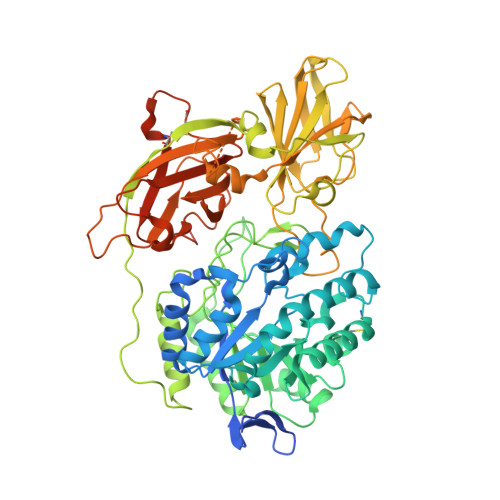 | |
UniProt & NIH Common Fund Data Resources | |||||
PHAROS: P16278 GTEx: ENSG00000170266 | |||||
Entity Groups | |||||
| Sequence Clusters | 30% Identity50% Identity70% Identity90% Identity95% Identity100% Identity | ||||
| UniProt Group | P16278 | ||||
Glycosylation | |||||
| Glycosylation Sites: 4 | Go to GlyGen: P16278-1 | ||||
Sequence AnnotationsExpand | |||||
Reference Sequence | |||||
| Ligands 5 Unique | |||||
|---|---|---|---|---|---|
| ID | Chains | Name / Formula / InChI Key | 2D Diagram | 3D Interactions | |
| N8V Download:Ideal Coordinates CCD File | DA [auth C], J [auth A], NA [auth D], T [auth B] | (1S,2S,3S,6R)-4-(hydroxymethyl)-6-(octylamino)cyclohex-4-ene-1,2,3-triol C15 H29 N O4 UPZUHYMBTUUPML-QPSCCSFWSA-N |  | ||
| NAG Download:Ideal Coordinates CCD File | AA [auth C] BA [auth C] E [auth A] F [auth A] G [auth A] | 2-acetamido-2-deoxy-beta-D-glucopyranose C8 H15 N O6 OVRNDRQMDRJTHS-FMDGEEDCSA-N |  | ||
| SO4 Download:Ideal Coordinates CCD File | EA [auth C] FA [auth C] K [auth A] L [auth A] OA [auth D] | SULFATE ION O4 S QAOWNCQODCNURD-UHFFFAOYSA-L |  | ||
| EDO Download:Ideal Coordinates CCD File | GA [auth C] HA [auth C] M [auth A] N [auth A] QA [auth D] | 1,2-ETHANEDIOL C2 H6 O2 LYCAIKOWRPUZTN-UHFFFAOYSA-N |  | ||
| CL Download:Ideal Coordinates CCD File | CA [auth C], I [auth A], MA [auth D], S [auth B] | CHLORIDE ION Cl VEXZGXHMUGYJMC-UHFFFAOYSA-M |  | ||
| Length ( Å ) | Angle ( ˚ ) |
|---|---|
| a = 94.933 | α = 90 |
| b = 115.915 | β = 92.25 |
| c = 140.51 | γ = 90 |
| Software Name | Purpose |
|---|---|
| REFMAC | refinement |
| PDB_EXTRACT | data extraction |
| HKL-2000 | data collection |
| HKL-2000 | data reduction |
| HKL-2000 | data scaling |
| MOLREP | phasing |


