3W9S

Crystal Structure Analysis of the N-terminal Receiver domain of Response Regulator PmrA


Experimental Data Snapshot

  • Method: X-RAY DIFFRACTION
  • Resolution: 1.70 Å
  • R-Value Free: 0.175 
  • R-Value Work: 0.165 
  • R-Value Observed: 0.166 

Starting Model: experimental
View more details

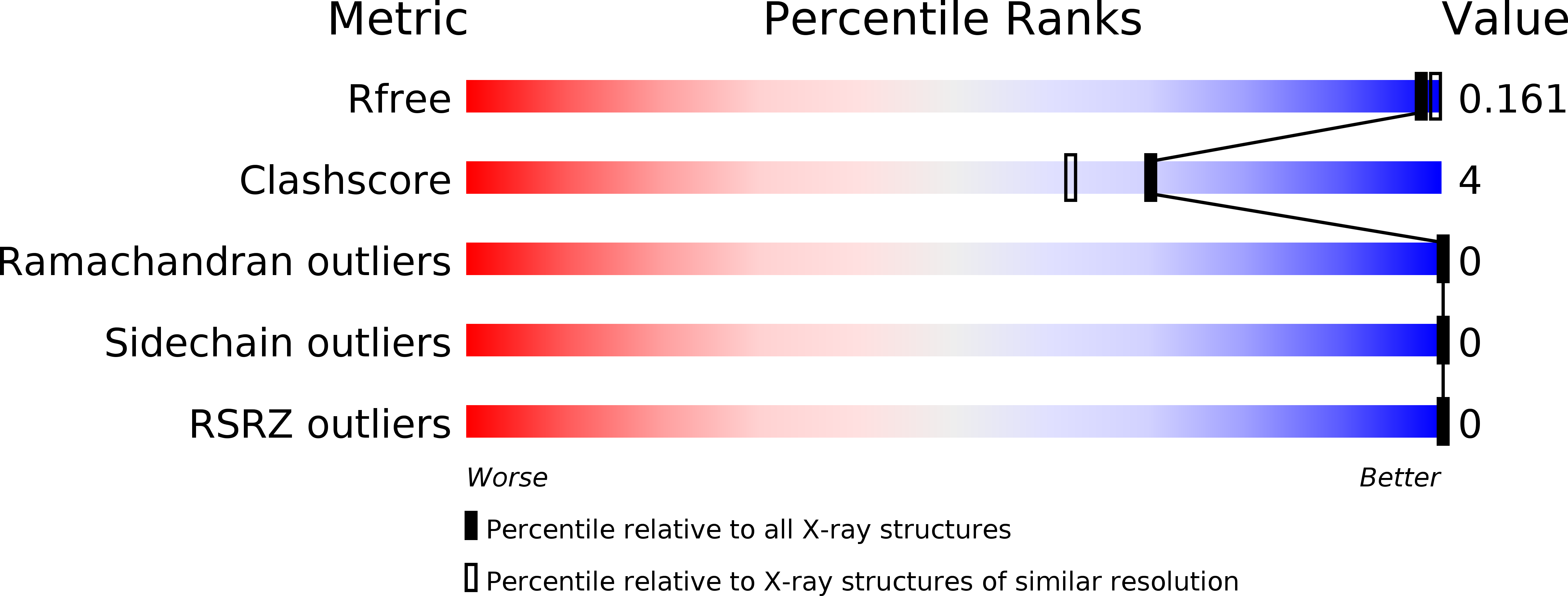
wwPDB Validation   3D Report Full Report


This is version 1.2 of the entry. See complete history


Literature

Structural Basis of a Physical Blockage Mechanism for the Interaction of Response Regulator PmrA with Connector Protein PmrD from Klebsiella Pneumoniae

Lou, S.C.Lou, Y.C.Rajasekaran, M.Chang, Y.W.Hsiao, C.D.Chen, C.

(2013) J Biol Chem 288: 25551-25561

  • DOI: https://doi.org/10.1074/jbc.M113.481978
  • Primary Citation of Related Structures:  
    3W9S

  • PubMed Abstract: 

    In bacteria, the two-component system is the most prevalent for sensing and transducing environmental signals into the cell. The PmrA-PmrB two-component system, responsible for sensing external stimuli of high Fe(3+) and mild acidic conditions, can control the genes involved in lipopolysaccharide modification and polymyxin resistance in pathogens. In Klebsiella pneumoniae, the small basic connector protein PmrD protects phospho-PmrA and prolongs the expression of PmrA-activated genes. We previously determined the phospho-PmrA recognition mode of PmrD. However, how PmrA interacts with PmrD and prevents its dephosphorylation remains unknown. To address this question, we solved the x-ray crystal structure of the N-terminal receiver domain of BeF3(-)-activated PmrA (PmrA(N)) at 1.70 Å. With this structure, we applied the data-driven docking method based on NMR chemical shift perturbation to generate the complex model of PmrD-PmrA(N), which was further validated by site-directed spin labeling experiments. In the complex model, PmrD may act as a blockade to prevent phosphatase from contacting with the phosphorylation site on PmrA.


  • Organizational Affiliation

    From the Institutes of Biomedical Sciences and.


Macromolecules
Find similar proteins by:  (by identity cutoff)  |  3D Structure
Entity ID: 1
MoleculeChains Sequence LengthOrganismDetailsImage
OmpR family response regulator in two-component regulatory system with BasS
A, B
131Klebsiella pneumoniae subsp. pneumoniae NTUH-K2044Mutation(s): 0 
Gene Names: KP1_1760
Entity Groups  
Sequence Clusters30% Identity50% Identity70% Identity90% Identity95% Identity100% Identity
Sequence Annotations
Expand
  • Reference Sequence
Experimental Data & Validation

Experimental Data

  • Method: X-RAY DIFFRACTION
  • Resolution: 1.70 Å
  • R-Value Free: 0.175 
  • R-Value Work: 0.165 
  • R-Value Observed: 0.166 
  • Space Group: P 41
Unit Cell:
Length ( Å )Angle ( ˚ )
a = 49.994α = 90
b = 49.994β = 90
c = 145.859γ = 90
Software Package:
Software NamePurpose
HKL-2000data collection
SOLVEphasing
CNSrefinement
HKL-2000data reduction
HKL-2000data scaling
PHENIXrefinement

Structure Validation

View Full Validation Report


Entry History 

Deposition Data

Revision History  (Full details and data files)

  • Version 1.0: 2013-07-24
    Type: Initial release
  • Version 1.1: 2013-09-18
    Changes: Database references
  • Version 1.2: 2023-11-08
    Changes: Data collection, Database references, Derived calculations, Refinement description