3W9C | pdb_00003w9c

Crystal structure of the electron transfer complex of cytochrome p450cam with putidaredoxin


Experimental Data Snapshot

  • Method: X-RAY DIFFRACTION
  • Resolution: 2.50 Å
  • R-Value Free: 
    0.251 (Depositor), 0.250 (DCC) 
  • R-Value Work: 
    0.185 (Depositor), 0.185 (DCC) 
  • R-Value Observed: 
    0.188 (Depositor) 

Starting Models: experimental
View more details

wwPDB Validation 3D Report Full Report

Validation slider image for 3W9C

Ligand Structure Quality Assessment 


This is version 1.2 of the entry. See complete history

Literature

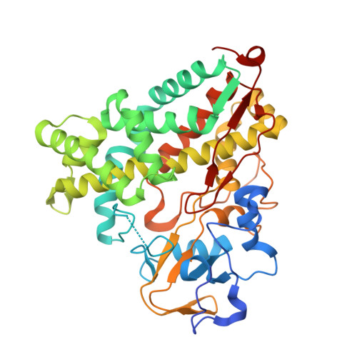
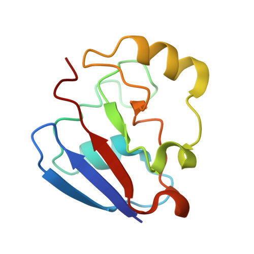
The structure of the cytochrome p450cam-putidaredoxin complex determined by paramagnetic NMR spectroscopy and crystallography.

Hiruma, Y.Hass, M.A.Kikui, Y.Liu, W.M.Olmez, B.Skinner, S.P.Blok, A.Kloosterman, A.Koteishi, H.Lohr, F.Schwalbe, H.Nojiri, M.Ubbink, M.

(2013) J Mol Biology 425: 4353-4365

  • DOI: https://doi.org/10.1016/j.jmb.2013.07.006
  • Primary Citation Related Structures: 
    2M56, 3W9C

  • PubMed Abstract: 

    Cytochrome P450cam catalyzes the hydroxylation of camphor in a complex process involving two electron transfers (ETs) from the iron-sulfur protein putidaredoxin. The enzymatic control of the successive steps of catalysis is critical for a highly efficient reaction. The injection of the successive electrons is part of the control system. To understand the molecular interactions between putidaredoxin and cytochrome P450cam, we determined the structure of the complex both in solution and in the crystal state. Paramagnetic NMR spectroscopy using lanthanide tags yielded 446 structural restraints that were used to determine the solution structure. An ensemble of 10 structures with an RMSD of 1.3Å was obtained. The crystal structure of the complex was solved, showing a position of putidaredoxin that is identical with the one in the solution structure. The NMR data further demonstrate the presence of a minor state or set of states of the complex in solution, which is attributed to the presence of an encounter complex. The structure of the major state shows a small binding interface and a metal-to-metal distance of 16Å, with two pathways that provide strong electronic coupling of the redox centers. The interpretation of these results is discussed in the context of ET. The structure indicates that the ET rate can be much faster than the reported value, suggesting that the process may be gated.


  • Organizational Affiliation
    • Leiden Institute of Chemistry, Gorlaeus Laboratories, Leiden University, Einsteinweg 55, 2333 CC Leiden, The Netherlands. Electronic address: hirumay@chem.leidenuniv.nl.

Macromolecule Content 

  • Total Structure Weight: 59.53 kDa 
  • Atom Count: 4,078 
  • Modeled Residue Count: 507 
  • Deposited Residue Count: 524 
  • Unique protein chains: 2

Macromolecules

Find similar proteins by:|  3D Structure
Entity ID: 1
MoleculeChains  Sequence LengthOrganismDetailsImage
Camphor 5-monooxygenase416Pseudomonas putidaMutation(s): 3 
Gene Names: camCcyp101
EC: 1.14.15.1
UniProt
Find proteins for P00183 (Pseudomonas putida)
Explore P00183 
Go to UniProtKB:  P00183
Entity Groups
Sequence Clusters30% Identity50% Identity70% Identity90% Identity95% Identity100% Identity
UniProt GroupP00183
Sequence Annotations
Expand
Reference Sequence
Find similar proteins by:|  3D Structure
Entity ID: 2
MoleculeChains  Sequence LengthOrganismDetailsImage
Putidaredoxin108Pseudomonas putidaMutation(s): 1 
Gene Names: camB
UniProt
Find proteins for P00259 (Pseudomonas putida)
Explore P00259 
Go to UniProtKB:  P00259
Entity Groups
Sequence Clusters30% Identity50% Identity70% Identity90% Identity95% Identity100% Identity
UniProt GroupP00259
Sequence Annotations
Expand
Reference Sequence

Small Molecules

Ligands 4 Unique
IDChains Name / Formula / InChI Key2D Diagram3D Interactions
HEM

Query on HEM


Download:Ideal Coordinates CCD File
C [auth A]PROTOPORPHYRIN IX CONTAINING FE
C34 H32 Fe N4 O4
KABFMIBPWCXCRK-RGGAHWMASA-L
FES

Query on FES


Download:Ideal Coordinates CCD File
H [auth B]FE2/S2 (INORGANIC) CLUSTER
Fe2 S2
NIXDOXVAJZFRNF-UHFFFAOYSA-N
SO4

Query on SO4


Download:Ideal Coordinates CCD File
D [auth A],
E [auth A],
F [auth A],
I [auth B]
SULFATE ION
O4 S
QAOWNCQODCNURD-UHFFFAOYSA-L
GOL

Query on GOL


Download:Ideal Coordinates CCD File
G [auth A]GLYCEROL
C3 H8 O3
PEDCQBHIVMGVHV-UHFFFAOYSA-N

Experimental Data & Validation

Experimental Data

  • Method: X-RAY DIFFRACTION
  • Resolution: 2.50 Å
  • R-Value Free:  0.251 (Depositor), 0.250 (DCC) 
  • R-Value Work:  0.185 (Depositor), 0.185 (DCC) 
  • R-Value Observed: 0.188 (Depositor) 
Space Group: C 1 2 1
Unit Cell:
Length ( Å )Angle ( ˚ )
a = 101.717α = 90
b = 77.991β = 95.57
c = 60.016γ = 90
Software Package:
Software NamePurpose
HKL-2000data collection
MOLREPphasing
REFMACrefinement
HKL-2000data reduction
SCALEPACKdata scaling

Structure Validation

View Full Validation Report


Ligand Structure Quality Assessment 


Entry History 

Deposition Data

Revision History  (Full details and data files)

  • Version 1.0: 2013-08-21
    Type: Initial release
  • Version 1.1: 2022-08-24
    Changes: Database references, Derived calculations
  • Version 1.2: 2023-11-08
    Changes: Data collection, Refinement description