3VX4 | pdb_00003vx4

Crystal Structure of the Nucleotide-Binding Domain of S. mutans ComA, a Bifunctional ATP-binding Cassette Transporter Involved in the Quorum-sensing Pathway


Experimental Data Snapshot

  • Method: X-RAY DIFFRACTION
  • Resolution: 2.69 Å
  • R-Value Free: 
    0.248 (Depositor), 0.248 (DCC) 
  • R-Value Work: 
    0.191 (Depositor), 0.191 (DCC) 
  • R-Value Observed: 
    0.194 (Depositor) 

Starting Model: experimental
View more details

wwPDB Validation 3D Report Full Report

Validation slider image for 3VX4

Ligand Structure Quality Assessment 


This is version 1.2 of the entry. See complete history

Literature

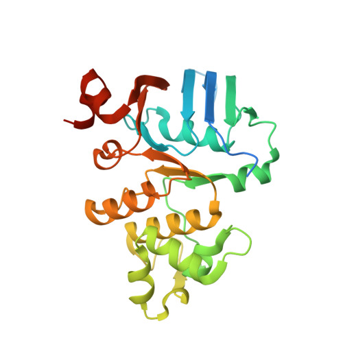
Boundary of the Nucleotide-Binding Domain of Streptococcus ComA Based on Functional and Structural Analysis

Ishii, S.Yano, T.Okamoto, A.Murakawa, T.Hayashi, H.

(2013) Biochemistry 52: 2545-2555

  • DOI: https://doi.org/10.1021/bi3017069
  • Primary Citation Related Structures: 
    3VX4

  • PubMed Abstract: 

    The ATP-binding cassette (ABC) transporter ComA is a key molecule essential for the first step of the quorum-sensing system of Streptococcus. The nucleotide binding domains (NBD) of Streptococcus mutans ComA with different N termini, NBD1 (amino acid residues 495-760), NBD2 (517-760), and NBD3 (528-760), were expressed, purified, and characterized. The shortest NBD3 corresponds to the region commonly defined as NBD in the database searches of ABC transporters. A kinetic analysis showed that the extra N-terminal region conferred a significantly higher ATP hydrolytic activity on the NBD at a neutral pH. Gel-filtration, X-ray crystallography, and mutational analyses suggest that at least four to five residues beyond the N-terminal boundary of NBD3 indeed participate in stabilizing the protein scaffold of the domain structure, thereby facilitating the ATP-dependent dimerization of NBD which is a prerequisite to the catalysis. These findings, together with the presence of a highly conserved glycine residue in this region, support the redefinition of the N-terminal boundary of the NBD of these types of ABC exporters.


  • Organizational Affiliation
    • Department of Biochemistry, Faculty of Nursing, Osaka Medical College, Osaka 569-8686, Japan. med002@art.osaka-med.ac.jp

Macromolecule Content 

  • Total Structure Weight: 61.76 kDa 
  • Atom Count: 3,914 
  • Modeled Residue Count: 479 
  • Deposited Residue Count: 546 
  • Unique protein chains: 1

Macromolecules

Find similar proteins by:|  3D Structure
Entity ID: 1
MoleculeChains  Sequence LengthOrganismDetailsImage
Putative ABC transporter, ATP-binding protein ComAA,
B [auth D]
273Streptococcus mutans UA159Mutation(s): 1 
Gene Names: SMU_286
UniProt
Find proteins for Q8DW05 (Streptococcus mutans serotype c (strain ATCC 700610 / UA159))
Explore Q8DW05 
Go to UniProtKB:  Q8DW05
Entity Groups
Sequence Clusters30% Identity50% Identity70% Identity90% Identity95% Identity100% Identity
UniProt GroupQ8DW05
Sequence Annotations
Expand
Reference Sequence

Experimental Data & Validation

Experimental Data

  • Method: X-RAY DIFFRACTION
  • Resolution: 2.69 Å
  • R-Value Free:  0.248 (Depositor), 0.248 (DCC) 
  • R-Value Work:  0.191 (Depositor), 0.191 (DCC) 
  • R-Value Observed: 0.194 (Depositor) 
Space Group: P 62
Unit Cell:
Length ( Å )Angle ( ˚ )
a = 129.611α = 90
b = 129.611β = 90
c = 72.425γ = 120
Software Package:
Software NamePurpose
HKL-2000data collection
MOLREPphasing
REFMACrefinement
HKL-2000data reduction
HKL-2000data scaling

Structure Validation

View Full Validation Report


Ligand Structure Quality Assessment 


Entry History 

Deposition Data

Revision History  (Full details and data files)

  • Version 1.0: 2013-04-17
    Type: Initial release
  • Version 1.1: 2013-06-05
    Changes: Database references
  • Version 1.2: 2023-11-08
    Changes: Data collection, Database references, Derived calculations, Refinement description