3VV7 | pdb_00003vv7

Crystal Structure of beta secetase in complex with 2-amino-6-((1S,2R)-2-(3'-methoxybiphenyl-3-yl)cyclopropyl)-3-methylpyrimidin-4(3H)-one


Experimental Data Snapshot

  • Method: X-RAY DIFFRACTION
  • Resolution: 2.10 Å
  • R-Value Free: 
    0.280 (Depositor), 0.279 (DCC) 
  • R-Value Work: 
    0.242 (Depositor), 0.243 (DCC) 
  • R-Value Observed: 
    0.244 (Depositor) 

wwPDB Validation 3D Report Full Report

Validation slider image for 3VV7

Ligand Structure Quality Assessment 


This is version 1.2 of the entry. See complete history

Literature

Conformational restriction approach to beta-secretase (BACE1) inhibitors: effect of a cyclopropane ring to induce an alternative binding mode.

Yonezawa, S.Yamamoto, T.Yamakawa, H.Muto, C.Hosono, M.Hattori, K.Higashino, K.Yutsudo, T.Iwamoto, H.Kondo, Y.Sakagami, M.Togame, H.Tanaka, Y.Nakano, T.Takemoto, H.Arisawa, M.Shuto, S.

(2012) J Med Chem 55: 8838-8858

  • DOI: https://doi.org/10.1021/jm3011405
  • Primary Citation Related Structures: 
    3VV6, 3VV7, 3VV8

  • PubMed Abstract: 

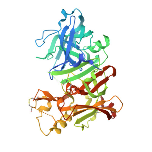
    Improvement of a drug's binding activity using the conformational restriction approach with sp³ hybridized carbon is becoming a key strategy in drug discovery. We applied this approach to BACE1 inhibitors and designed four stereoisomeric cyclopropane compounds in which the ethylene linker of a known amidine-type inhibitor 2 was replaced with chiral cyclopropane rings. The synthesis and biologic evaluation of these compounds revealed that the cis-(1S,2R) isomer 6 exhibited the most potent BACE1 inhibitory activity among them. X-ray structure analysis of the complex of 6 and BACE1 revealed that its unique binding mode is due to the apparent CH-π interaction between the rigid cyclopropane ring and the Tyr71 side chain. A derivatization study using 6 as a lead molecule led to the development of highly potent inhibitors in which the structure-activity relationship as well as the binding mode of the compounds clearly differ from those of known amidine-type inhibitors.


  • Organizational Affiliation
    • Shionogi Innovation Center for Drug Discovery, Shionogi & Co., Ltd., Kita-21 Nishi-11 Kita-ku, Sapporo 001-0021, Japan. shu@pharm.hokudai.ac.jp

Macromolecule Content 

  • Total Structure Weight: 47.69 kDa 
  • Atom Count: 3,065 
  • Modeled Residue Count: 361 
  • Deposited Residue Count: 416 
  • Unique protein chains: 1

Macromolecules

Find similar proteins by:|  3D Structure
Entity ID: 1
MoleculeChains  Sequence LengthOrganismDetailsImage
Beta-secretase 1416Homo sapiensMutation(s): 0 
Gene Names: BACE1
EC: 3.4.23.46
UniProt & NIH Common Fund Data Resources
Find proteins for P56817 (Homo sapiens)
Explore P56817 
Go to UniProtKB:  P56817
PHAROS:  P56817
GTEx:  ENSG00000186318 
Entity Groups
Sequence Clusters30% Identity50% Identity70% Identity90% Identity95% Identity100% Identity
UniProt GroupP56817
Sequence Annotations
Expand
Reference Sequence

Small Molecules

Ligands 3 Unique
IDChains Name / Formula / InChI Key2D Diagram3D Interactions
0B1

Query on 0B1


Download:Ideal Coordinates CCD File
J [auth A]2-amino-6-[(1S,2R)-2-(3'-methoxybiphenyl-3-yl)cyclopropyl]-3-methylpyrimidin-4(3H)-one
C21 H21 N3 O2
QECXNBWGUGUPSI-ROUUACIJSA-N
IOD

Query on IOD


Download:Ideal Coordinates CCD File
B [auth A]
C [auth A]
D [auth A]
E [auth A]
F [auth A]
B [auth A],
C [auth A],
D [auth A],
E [auth A],
F [auth A],
G [auth A]
IODIDE ION
I
XMBWDFGMSWQBCA-UHFFFAOYSA-M
GOL

Query on GOL


Download:Ideal Coordinates CCD File
H [auth A],
I [auth A]
GLYCEROL
C3 H8 O3
PEDCQBHIVMGVHV-UHFFFAOYSA-N

Experimental Data & Validation

Experimental Data

  • Method: X-RAY DIFFRACTION
  • Resolution: 2.10 Å
  • R-Value Free:  0.280 (Depositor), 0.279 (DCC) 
  • R-Value Work:  0.242 (Depositor), 0.243 (DCC) 
  • R-Value Observed: 0.244 (Depositor) 
Space Group: P 61 2 2
Unit Cell:
Length ( Å )Angle ( ˚ )
a = 103.681α = 90
b = 103.681β = 90
c = 169.006γ = 120
Software Package:
Software NamePurpose
DENZOdata reduction
SCALEPACKdata scaling
MOLREPphasing
REFMACrefinement
PDB_EXTRACTdata extraction
HKL-2000data collection
HKL-2000data reduction
HKL-2000data scaling

Structure Validation

View Full Validation Report


Ligand Structure Quality Assessment 


Entry History 

Revision History  (Full details and data files)

  • Version 1.0: 2012-10-24
    Type: Initial release
  • Version 1.1: 2013-09-04
    Changes: Database references
  • Version 1.2: 2024-11-20
    Changes: Data collection, Database references, Derived calculations, Structure summary