3VTE | pdb_00003vte

Crystal structure of tetrahydrocannabinolic acid synthase from Cannabis sativa


Experimental Data Snapshot

  • Method: X-RAY DIFFRACTION
  • Resolution: 2.75 Å
  • R-Value Free: 
    0.250 (Depositor), 0.242 (DCC) 
  • R-Value Work: 
    0.198 (Depositor), 0.194 (DCC) 
  • R-Value Observed: 
    0.201 (Depositor) 

Starting Model: experimental
View more details

wwPDB Validation 3D Report Full Report

Validation slider image for 3VTE

Ligand Structure Quality Assessment 


This is version 1.4 of the entry. See complete history

Literature

Structure and function of 1-tetrahydrocannabinolic acid (THCA) synthase, the enzyme controlling the psychoactivity of Cannabis sativa

Shoyama, Y.Tamada, T.Kurihara, K.Takeuchi, A.Taura, F.Arai, S.Blaber, M.Shoyama, Y.Morimoto, S.Kuroki, R.

(2012) J Mol Biology 423: 96-105

  • DOI: https://doi.org/10.1016/j.jmb.2012.06.030
  • Primary Citation Related Structures: 
    3VTE

  • PubMed Abstract: 

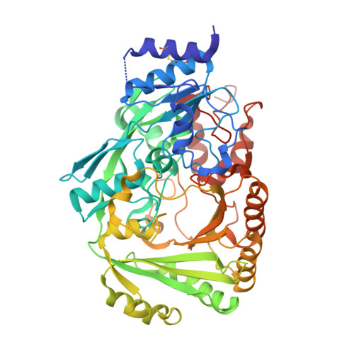
    ∆1-Tetrahydrocannabinolic acid (THCA) synthase catalyzes the oxidative cyclization of cannabigerolic acid (CBGA) into THCA, the precursor of the primary psychoactive agent ∆1-tetrahydrocannabinol in Cannabis sativa. The enzyme was overproduced in insect cells, purified, and crystallized in order to investigate the structure-function relationship of THCA synthase, and the tertiary structure was determined to 2.75Å resolution by X-ray crystallography (R(cryst)=19.9%). The THCA synthase enzyme is a member of the p-cresol methyl-hydroxylase superfamily, and the tertiary structure is divided into two domains (domains I and II), with a flavin adenine dinucleotide coenzyme positioned between each domain and covalently bound to His114 and Cys176 (located in domain I). The catalysis of THCA synthesis involves a hydride transfer from C3 of CBGA to N5 of flavin adenine dinucleotide and the deprotonation of O6' of CBGA. The ionized residues in the active site of THCA synthase were investigated by mutational analysis and X-ray structure. Mutational analysis indicates that the reaction does not involve the carboxyl group of Glu442 that was identified as the catalytic base in the related berberine bridge enzyme but instead involves the hydroxyl group of Tyr484. Mutations at the active-site residues His292 and Tyr417 resulted in a decrease in, but not elimination of, the enzymatic activity of THCA synthase, suggesting a key role for these residues in substrate binding and not direct catalysis.


  • Organizational Affiliation
    • Quantum Beam Science Directorate, Japan Atomic Energy Agency, 2-4 Shirakata-Shirane, Tokai, Ibaraki 319-1195, Japan. yoshinari_shoyama@nmp.co.jp

Macromolecule Content 

  • Total Structure Weight: 60.86 kDa 
  • Atom Count: 4,211 
  • Modeled Residue Count: 502 
  • Deposited Residue Count: 518 
  • Unique protein chains: 1

Macromolecules

Find similar proteins by:|  3D Structure
Entity ID: 1
MoleculeChains  Sequence LengthOrganismDetailsImage
Tetrahydrocannabinolic acid synthase518Cannabis sativaMutation(s): 0 
EC: 1.21.3.7
UniProt
Find proteins for Q8GTB6 (Cannabis sativa)
Explore Q8GTB6 
Go to UniProtKB:  Q8GTB6
Entity Groups
Sequence Clusters30% Identity50% Identity70% Identity90% Identity95% Identity100% Identity
UniProt GroupQ8GTB6
Glycosylation
Glycosylation Sites: 6
Sequence Annotations
Expand
Reference Sequence

Small Molecules

Ligands 2 Unique
IDChains Name / Formula / InChI Key2D Diagram3D Interactions
FAD

Query on FAD


Download:Ideal Coordinates CCD File
H [auth A]FLAVIN-ADENINE DINUCLEOTIDE
C27 H33 N9 O15 P2
VWWQXMAJTJZDQX-UYBVJOGSSA-N
NAG

Query on NAG


Download:Ideal Coordinates CCD File
B [auth A]
C [auth A]
D [auth A]
E [auth A]
F [auth A]
B [auth A],
C [auth A],
D [auth A],
E [auth A],
F [auth A],
G [auth A]
2-acetamido-2-deoxy-beta-D-glucopyranose
C8 H15 N O6
OVRNDRQMDRJTHS-FMDGEEDCSA-N

Experimental Data & Validation

Experimental Data

  • Method: X-RAY DIFFRACTION
  • Resolution: 2.75 Å
  • R-Value Free:  0.250 (Depositor), 0.242 (DCC) 
  • R-Value Work:  0.198 (Depositor), 0.194 (DCC) 
  • R-Value Observed: 0.201 (Depositor) 
Space Group: P 4 3 2
Unit Cell:
Length ( Å )Angle ( ˚ )
a = 177.946α = 90
b = 177.946β = 90
c = 177.946γ = 90
Software Package:
Software NamePurpose
ADSCdata collection
PHASERphasing
REFMACrefinement
HKL-2000data reduction
HKL-2000data scaling

Structure Validation

View Full Validation Report


Ligand Structure Quality Assessment 


Entry History 

Deposition Data

Revision History  (Full details and data files)

  • Version 1.0: 2012-07-25
    Type: Initial release
  • Version 1.1: 2014-04-30
    Changes: Database references
  • Version 1.2: 2020-07-29
    Type: Remediation
    Reason: Carbohydrate remediation
    Changes: Data collection, Derived calculations, Structure summary
  • Version 1.3: 2023-11-08
    Changes: Data collection, Database references, Refinement description, Structure summary
  • Version 1.4: 2024-10-30
    Changes: Structure summary