3VRS | pdb_00003vrs

Crystal structure of fluoride riboswitch, soaked in Mn2+


Experimental Data Snapshot

  • Method: X-RAY DIFFRACTION
  • Resolution: 2.60 Å
  • R-Value Free: 
    0.262 (Depositor), 0.243 (DCC) 
  • R-Value Work: 
    0.220 (Depositor), 0.219 (DCC) 
  • R-Value Observed: 
    0.224 (Depositor) 

Starting Model: experimental
View more details

wwPDB Validation 3D Report Full Report

Validation slider image for 3VRS

This is version 1.3 of the entry. See complete history

Literature

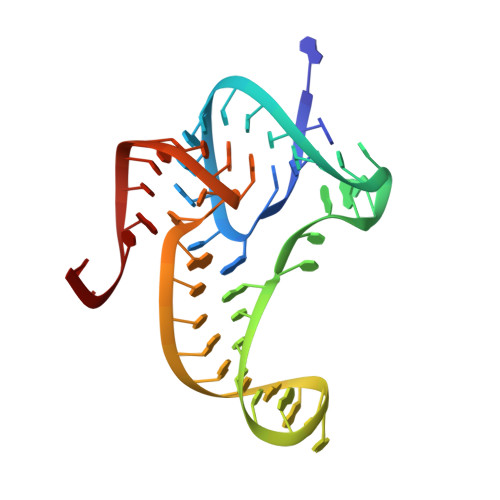
Fluoride ion encapsulation by Mg2+ ions and phosphates in a fluoride riboswitch.

Ren, A.Rajashankar, K.R.Patel, D.J.

(2012) Nature 486: 85-89

  • DOI: https://doi.org/10.1038/nature11152
  • Primary Citation Related Structures: 
    3VRS, 4EN5, 4ENA, 4ENB, 4ENC

  • PubMed Abstract: 

    Significant advances in our understanding of RNA architecture, folding and recognition have emerged from structure-function studies on riboswitches, non-coding RNAs whose sensing domains bind small ligands and whose adjacent expression platforms contain RNA elements involved in the control of gene regulation. We now report on the ligand-bound structure of the Thermotoga petrophila fluoride riboswitch, which adopts a higher-order RNA architecture stabilized by pseudoknot and long-range reversed Watson-Crick and Hoogsteen A•U pair formation. The bound fluoride ion is encapsulated within the junctional architecture, anchored in place through direct coordination to three Mg(2+) ions, which in turn are octahedrally coordinated to water molecules and five inwardly pointing backbone phosphates. Our structure of the fluoride riboswitch in the bound state shows how RNA can form a binding pocket selective for fluoride, while discriminating against larger halide ions. The T. petrophila fluoride riboswitch probably functions in gene regulation through a transcription termination mechanism.


  • Organizational Affiliation
    • Structural Biology Program, Memorial Sloan-Kettering Center, New York, New York 10065, USA.

Macromolecule Content 

  • Total Structure Weight: 17.33 kDa 
  • Atom Count: 1,139 
  • Modeled Residue Count: 52 
  • Deposited Residue Count: 52 
  • Unique nucleic acid chains: 1

Macromolecules

Find similar nucleic acids by:  (by identity cutoff) 
Entity ID: 1
MoleculeChains LengthOrganismImage
Fluoride riboswitch52Thermotoga petrophila
Sequence Annotations
Expand
Reference Sequence

Small Molecules

Ligands 3 Unique
IDChains Name / Formula / InChI Key2D Diagram3D Interactions
MN

Query on MN


Download:Ideal Coordinates CCD File
B [auth A]
C [auth A]
D [auth A]
E [auth A]
F [auth A]
B [auth A],
C [auth A],
D [auth A],
E [auth A],
F [auth A],
G [auth A],
H [auth A],
I [auth A],
J [auth A]
MANGANESE (II) ION
Mn
WAEMQWOKJMHJLA-UHFFFAOYSA-N
K

Query on K


Download:Ideal Coordinates CCD File
K [auth A]POTASSIUM ION
K
NPYPAHLBTDXSSS-UHFFFAOYSA-N
F

Query on F


Download:Ideal Coordinates CCD File
L [auth A]FLUORIDE ION
F
KRHYYFGTRYWZRS-UHFFFAOYSA-M

Experimental Data & Validation

Experimental Data

  • Method: X-RAY DIFFRACTION
  • Resolution: 2.60 Å
  • R-Value Free:  0.262 (Depositor), 0.243 (DCC) 
  • R-Value Work:  0.220 (Depositor), 0.219 (DCC) 
  • R-Value Observed: 0.224 (Depositor) 
Space Group: P 21 21 2
Unit Cell:
Length ( Å )Angle ( ˚ )
a = 57.846α = 90
b = 77.203β = 90
c = 42.094γ = 90
Software Package:
Software NamePurpose
ADSCdata collection
PHENIXmodel building
PHENIXrefinement
HKL-2000data reduction
HKL-2000data scaling
PHENIXphasing

Structure Validation

View Full Validation Report


Entry History 

Deposition Data

Revision History  (Full details and data files)

  • Version 1.0: 2012-05-09
    Type: Initial release
  • Version 1.1: 2012-06-20
    Changes: Database references
  • Version 1.2: 2013-02-13
    Changes: Structure summary
  • Version 1.3: 2023-09-13
    Changes: Data collection, Database references, Derived calculations, Refinement description