3VNW | pdb_00003vnw

Crystal structure of cytochrome c552 from Thermus thermophilus at pH 5.44


Experimental Data Snapshot

  • Method: X-RAY DIFFRACTION
  • Resolution: 1.97 Å
  • R-Value Free: 
    0.232 (Depositor), 0.230 (DCC) 
  • R-Value Work: 
    0.213 (Depositor), 0.211 (DCC) 
  • R-Value Observed: 
    0.214 (Depositor) 

Starting Model: experimental
View more details

wwPDB Validation 3D Report Full Report

Validation slider image for 3VNW

Ligand Structure Quality Assessment 


This is version 1.2 of the entry. See complete history

Literature

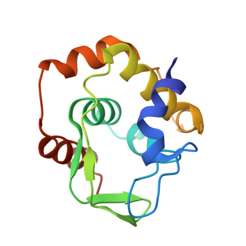
Snapshots of a protein folding intermediate

Yamada, S.Bouley-Ford, N.D.Keller, G.E.Ford, W.C.Gray, H.B.Winkler, J.R.

(2013) Proc Natl Acad Sci U S A 110: 1606-1610

  • DOI: https://doi.org/10.1073/pnas.1221832110
  • Primary Citation Related Structures: 
    3VNW

  • PubMed Abstract: 

    We have investigated the folding dynamics of Thermus thermophilus cytochrome c(552) by time-resolved fluorescence energy transfer between the heme and each of seven site-specific fluorescent probes. We have found both an equilibrium unfolding intermediate and a distinct refolding intermediate from kinetics studies. Depending on the protein region monitored, we observed either two-state or three-state denaturation transitions. The unfolding intermediate associated with three-state folding exhibited native contacts in β-sheet and C-terminal helix regions. We probed the formation of a refolding intermediate by time-resolved fluorescence energy transfer between residue 110 and the heme using a continuous flow mixer. The intermediate ensemble, a heterogeneous mixture of compact and extended polypeptides, forms in a millisecond, substantially slower than the ∼100-μs formation of a burst-phase intermediate in cytochrome c. The surprising finding is that, unlike for cytochrome c, there is an observable folding intermediate, but no microsecond burst phase in the folding kinetics of the structurally related thermostable protein.


  • Organizational Affiliation
    • Beckman Institute, California Institute of Technology, Pasadena, CA 91125, USA.

Macromolecule Content 

  • Total Structure Weight: 14.61 kDa 
  • Atom Count: 1,098 
  • Modeled Residue Count: 129 
  • Deposited Residue Count: 129 
  • Unique protein chains: 1

Macromolecules

Find similar proteins by:|  3D Structure
Entity ID: 1
MoleculeChains  Sequence LengthOrganismDetailsImage
Cytochrome c-552129Thermus thermophilusMutation(s): 0 
Gene Names: cycA
UniProt
Find proteins for P04164 (Thermus thermophilus)
Explore P04164 
Go to UniProtKB:  P04164
Entity Groups
Sequence Clusters30% Identity50% Identity70% Identity90% Identity95% Identity100% Identity
UniProt GroupP04164
Sequence Annotations
Expand
Reference Sequence

Small Molecules

Ligands 1 Unique
IDChains Name / Formula / InChI Key2D Diagram3D Interactions
HEM

Query on HEM


Download:Ideal Coordinates CCD File
B [auth A]PROTOPORPHYRIN IX CONTAINING FE
C34 H32 Fe N4 O4
KABFMIBPWCXCRK-RGGAHWMASA-L

Experimental Data & Validation

Experimental Data

  • Method: X-RAY DIFFRACTION
  • Resolution: 1.97 Å
  • R-Value Free:  0.232 (Depositor), 0.230 (DCC) 
  • R-Value Work:  0.213 (Depositor), 0.211 (DCC) 
  • R-Value Observed: 0.214 (Depositor) 
Space Group: P 65
Unit Cell:
Length ( Å )Angle ( ˚ )
a = 86.98α = 90
b = 86.98β = 90
c = 31.82γ = 120
Software Package:
Software NamePurpose
CrystalCleardata collection
MOLREPphasing
REFMACrefinement
CrystalCleardata reduction
CrystalCleardata scaling

Structure Validation

View Full Validation Report


Ligand Structure Quality Assessment 


Entry History 

Deposition Data

Revision History  (Full details and data files)

  • Version 1.0: 2013-01-23
    Type: Initial release
  • Version 1.1: 2013-02-06
    Changes: Database references
  • Version 1.2: 2023-11-08
    Changes: Data collection, Database references, Derived calculations, Refinement description