3VMX | pdb_00003vmx

Crystal Structure of a parallel coiled-coil dimerization domain from the voltage-gated proton channel


Experimental Data Snapshot

  • Method: X-RAY DIFFRACTION
  • Resolution: 1.45 Å
  • R-Value Free: 
    0.214 (Depositor) 
  • R-Value Work: 
    0.179 (Depositor), 0.193 (DCC) 
  • R-Value Observed: 
    0.181 (Depositor) 

wwPDB Validation 3D Report Full Report

Validation slider image for 3VMX

This is version 1.2 of the entry. See complete history

Literature

The cytoplasmic coiled-coil mediates cooperative gating temperature sensitivity in the voltage-gated H(+) channel Hv1

Fujiwara, Y.Kurokawa, T.Takeshita, K.Kobayashi, M.Okochi, Y.Nakagawa, A.Okamura, Y.

(2012) Nat Commun 3: 816-816

  • DOI: https://doi.org/10.1038/ncomms1823
  • Primary Citation Related Structures: 
    3VMX

  • PubMed Abstract: 

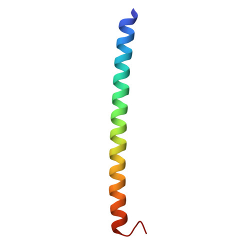
    Hv1/VSOP is a dimeric voltage-gated H(+) channel in which the gating of one subunit is reportedly coupled to that of the other subunit within the dimer. The molecular basis for dimer formation and intersubunit coupling, however, remains unknown. Here we show that the carboxy terminus ends downstream of the S4 voltage-sensor helix twist in a dimer coiled-coil architecture, which mediates cooperative gating. We also show that the temperature-dependent activation of H(+) current through Hv1/VSOP is regulated by thermostability of the coiled-coil domain, and that this regulation is altered by mutation of the linker between S4 and the coiled-coil. Cooperative gating within the dimer is also dependent on the linker structure, which circular dichroism spectrum analysis suggests is α-helical. Our results indicate that the cytoplasmic coiled-coil strands form continuous α-helices with S4 and mediate cooperative gating to adjust the range of temperatures over which Hv1/VSOP operates.


  • Organizational Affiliation
    • Laboratory of Integrative Physiology, Department of Physiology, Graduate School and Faculty of Medicine, Osaka University, Yamadaoka 2-2, Suita, Osaka 565-0871, Japan. fujiwara@phys2.med.osaka-u.ac.jp

Macromolecule Content 

  • Total Structure Weight: 22.39 kDa 
  • Atom Count: 1,949 
  • Modeled Residue Count: 191 
  • Deposited Residue Count: 192 
  • Unique protein chains: 1

Macromolecules

Find similar proteins by:|  3D Structure
Entity ID: 1
MoleculeChains  Sequence LengthOrganismDetailsImage
Voltage-gated hydrogen channel 1A,
B [auth C],
C [auth B],
D
48Mus musculusMutation(s): 0 
Gene Names: BtsHvcn1Vsop
UniProt
Find proteins for Q3U2S8 (Mus musculus)
Explore Q3U2S8 
Go to UniProtKB:  Q3U2S8
Entity Groups
Sequence Clusters30% Identity50% Identity70% Identity90% Identity95% Identity100% Identity
UniProt GroupQ3U2S8
Sequence Annotations
Expand
Reference Sequence

Experimental Data & Validation

Experimental Data

  • Method: X-RAY DIFFRACTION
  • Resolution: 1.45 Å
  • R-Value Free:  0.214 (Depositor) 
  • R-Value Work:  0.179 (Depositor), 0.193 (DCC) 
  • R-Value Observed: 0.181 (Depositor) 
Space Group: P 21 21 21
Unit Cell:
Length ( Å )Angle ( ˚ )
a = 40.156α = 90
b = 54.01β = 90
c = 81.439γ = 90
Software Package:
Software NamePurpose
PHASERphasing
REFMACrefinement
HKL-2000data reduction
SCALEPACKdata scaling

Structure Validation

View Full Validation Report


Entry History 

Deposition Data

Revision History  (Full details and data files)

  • Version 1.0: 2012-05-30
    Type: Initial release
  • Version 1.1: 2013-06-26
    Changes: Database references
  • Version 1.2: 2024-03-20
    Changes: Data collection, Database references