3VM4 | pdb_00003vm4

Cytochrome P450SP alpha (CYP152B1) in complex with (R)-ibuprophen


Experimental Data Snapshot

  • Method: X-RAY DIFFRACTION
  • Resolution: 1.94 Å
  • R-Value Free: 
    0.186 (Depositor), 0.223 (DCC) 
  • R-Value Work: 
    0.158 (Depositor), 0.204 (DCC) 
  • R-Value Observed: 
    0.160 (Depositor) 

Starting Model: experimental
View more details

wwPDB Validation 3D Report Full Report

Validation slider image for 3VM4

Ligand Structure Quality Assessment 


This is version 1.2 of the entry. See complete history

Literature

Chiral-substrate-assisted stereoselective epoxidation catalyzed by H2O2-dependent cytochrome P450SP alpha

Fujishiro, T.Shoji, O.Kawakami, N.Watanabe, T.Sugimoto, H.Shiro, Y.Watanabe, Y.

(2012) Chem Asian J 7: 2286-2293

  • DOI: https://doi.org/10.1002/asia.201200250
  • Primary Citation Related Structures: 
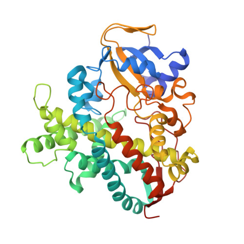
    3VM4

  • PubMed Abstract: 

    The stereoselective epoxidation of styrene was catalyzed by H(2) O(2) -dependent cytochrome P450(SPα) in the presence of carboxylic acids as decoy molecules. The stereoselectivity of styrene oxide could be altered by the nature of the decoy molecules. In particular, the chirality at the α-positions of the decoy molecules induced a clear difference in the chirality of the product: (R)-ibuprofen enhanced the formation of (S)-styrene oxide, whereas (S)-ibuprofen preferentially afforded (R)-styrene oxide. The crystal structure of an (R)-ibuprofen-bound cytochrome P450(SPα) (resolution 1.9 Å) revealed that the carboxylate group of (R)-ibuprofen served as an acid-base catalyst to initiate the epoxidation. A docking simulation of the binding of styrene in the active site of the (R)-ibuprofen-bound form suggested that the orientation of the vinyl group of styrene in the active site agreed with the formation of (S)-styrene oxide.


  • Organizational Affiliation
    • Department of Chemistry, Graduate School of Science, Nagoya University, Furo-cho, Chikusa-ku, Nagoya 464-8602, Japan.

Macromolecule Content 

  • Total Structure Weight: 46.68 kDa 
  • Atom Count: 3,614 
  • Modeled Residue Count: 407 
  • Deposited Residue Count: 407 
  • Unique protein chains: 1

Macromolecules

Find similar proteins by:|  3D Structure
Entity ID: 1
MoleculeChains  Sequence LengthOrganismDetailsImage
Fatty acid alpha-hydroxylase407Sphingomonas paucimobilisMutation(s): 0 
EC: 1.11.2.4
UniProt
Find proteins for O24782 (Sphingomonas paucimobilis)
Explore O24782 
Go to UniProtKB:  O24782
Entity Groups
Sequence Clusters30% Identity50% Identity70% Identity90% Identity95% Identity100% Identity
UniProt GroupO24782
Sequence Annotations
Expand
Reference Sequence

Experimental Data & Validation

Experimental Data

  • Method: X-RAY DIFFRACTION
  • Resolution: 1.94 Å
  • R-Value Free:  0.186 (Depositor), 0.223 (DCC) 
  • R-Value Work:  0.158 (Depositor), 0.204 (DCC) 
  • R-Value Observed: 0.160 (Depositor) 
Space Group: P 31 2 1
Unit Cell:
Length ( Å )Angle ( ˚ )
a = 94.39α = 90
b = 94.39β = 90
c = 112.863γ = 120
Software Package:
Software NamePurpose
DENZOdata reduction
SCALEPACKdata scaling
MOLREPphasing
REFMACrefinement
PDB_EXTRACTdata extraction
HKL-2000data collection

Structure Validation

View Full Validation Report


Ligand Structure Quality Assessment 


Entry History 

Deposition Data

Revision History  (Full details and data files)

  • Version 1.0: 2012-05-09
    Type: Initial release
  • Version 1.1: 2013-08-14
    Changes: Database references
  • Version 1.2: 2023-11-08
    Changes: Data collection, Database references, Derived calculations, Refinement description