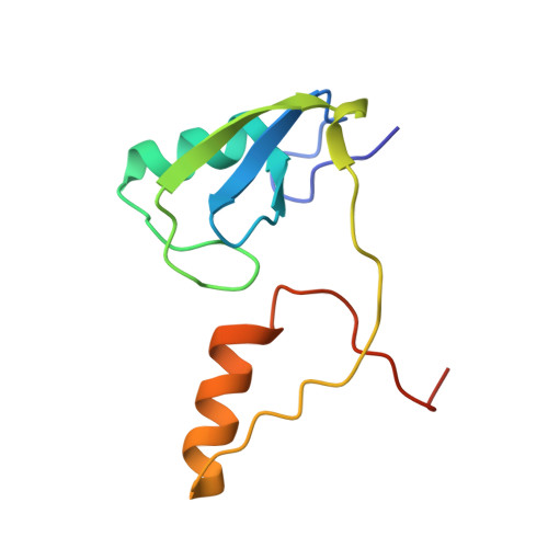
3VK6 | pdb_00003vk6

Crystal structure of a phosphotyrosine binding domain


Experimental Data Snapshot

  • Method: X-RAY DIFFRACTION
  • Resolution: 1.90 Å
  • R-Value Free: 
    0.242 (Depositor), 0.236 (DCC) 
  • R-Value Work: 
    0.219 (Depositor), 0.212 (DCC) 
  • R-Value Observed: 
    0.222 (Depositor) 

wwPDB Validation 3D Report Full Report

Validation slider image for 3VK6

This is version 1.2 of the entry. See complete history. 

Literature

Structure of a novel phosphotyrosine-binding domain in Hakai that targets E-cadherin

Mukherjee, M., Chow, S.Y., Yusoff, P., Seetharaman, J., Ng, C., Sinniah, S., Koh, X.W., Asgar, N.F., Li, D., Yim, D., Jackson, R.A., Yew, J., Qian, J., Iyu, A., Lim, Y.P., Zhou, X., Sze, S.K., Guy, G.R., Sivaraman, J.

(2012) EMBO J 31: 1308-1319

  • DOI: https://doi.org/10.1038/emboj.2011.496
  • Primary Citation Related Structures: 
    3VK6

  • PubMed Abstract: 

    Phosphotyrosine-binding domains, typified by the SH2 (Src homology 2) and PTB domains, are critical upstream components of signal transduction pathways. The E3 ubiquitin ligase Hakai targets tyrosine-phosphorylated E-cadherin via an uncharacterized domain. In this study, the crystal structure of Hakai (amino acids 106-206) revealed that it forms an atypical, zinc-coordinated homodimer by utilizing residues from the phosphotyrosine-binding domain of two Hakai monomers. Hakai dimerization allows the formation of a phosphotyrosine-binding pocket that recognizes specific phosphorylated tyrosines and flanking acidic amino acids of Src substrates, such as E-cadherin, cortactin and DOK1. NMR and mutational analysis identified the Hakai residues required for target binding within the binding pocket, now named the HYB domain. ZNF645 also possesses a HYB domain but demonstrates different target specificities. The HYB domain is structurally different from other phosphotyrosine-binding domains and is a potential drug target due to its novel structural features.


  • Organizational Affiliation: 
    • Department of Biological Sciences, National University of Singapore, Singapore.

Macromolecule Content 

  • Total Structure Weight: 11.75 kDa 
  • Atom Count: 828 
  • Modeled Residue Count: 96 
  • Deposited Residue Count: 101 
  • Unique protein chains: 1

Macromolecules

Find similar proteins by:|  3D Structure
Entity ID: 1
MoleculeChains  Sequence LengthOrganismDetailsImage
E3 ubiquitin-protein ligase Hakai101Mus musculusMutation(s): 0 
Gene Names: Cbll1, Hakai
EC: 6.3.2 (PDB Primary Data), 2.3.2.27 (UniProt)
UniProt
Find proteins for Q9JIY2 (Mus musculus)
Explore Q9JIY2 
Go to UniProtKB:  Q9JIY2
Entity Groups
Sequence Clusters30% Identity50% Identity70% Identity90% Identity95% Identity100% Identity
UniProt GroupQ9JIY2
Sequence Annotations
Expand
Reference Sequence

Experimental Data & Validation

Experimental Data

  • Method: X-RAY DIFFRACTION
  • Resolution: 1.90 Å
  • R-Value Free:  0.242 (Depositor), 0.236 (DCC) 
  • R-Value Work:  0.219 (Depositor), 0.212 (DCC) 
  • R-Value Observed: 0.222 (Depositor) 
Space Group: P 62 2 2
Unit Cell:
Length ( Å )Angle ( ˚ )
a = 64.657α = 90
b = 64.657β = 90
c = 121.035γ = 120
Software Package:
Software NamePurpose
HKL-2000data collection
SOLVEphasing
PHENIXrefinement
HKL-2000data reduction
HKL-2000data scaling

Structure Validation

View Full Validation Report


Entry History 

Deposition Data

Revision History  (Full details and data files)

  • Version 1.0: 2012-01-25
    Type: Initial release
  • Version 1.1: 2013-07-24
    Changes: Database references
  • Version 1.2: 2024-03-20
    Changes: Data collection, Database references, Derived calculations