3VJF | pdb_00003vjf

Crystal structure of de novo 4-helix bundle protein WA20


Experimental Data Snapshot

  • Method: X-RAY DIFFRACTION
  • Resolution: 2.20 Å
  • R-Value Free: 
    0.255 (Depositor), 0.265 (DCC) 
  • R-Value Work: 
    0.233 (Depositor), 0.257 (DCC) 
  • R-Value Observed: 
    0.234 (Depositor) 

wwPDB Validation 3D Report Full Report

Validation slider image for 3VJF

This is version 1.3 of the entry. See complete history

Literature

Domain-swapped dimeric structure of a stable and functional de novo four-helix bundle protein, WA20

Arai, R.Kobayashi, N.Kimura, A.Sato, T.Matsuo, K.Wang, A.F.Platt, J.M.Bradley, L.H.Hecht, M.H.

(2012) J Phys Chem B 116: 6789-6797

  • DOI: https://doi.org/10.1021/jp212438h
  • Primary Citation Related Structures: 
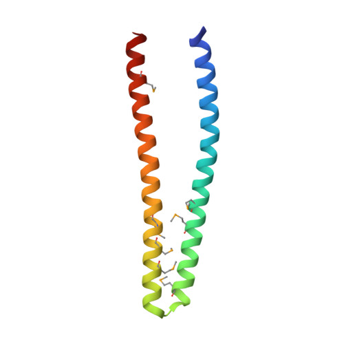
    3VJF

  • PubMed Abstract: 

    To probe the potential for activity in unevolved amino acid sequence space, we created a third generation combinatorial library of de novo four-helix bundle proteins. The "artificial superfamily" of helical bundles was designed using binary patterning of polar and nonpolar residues, and expressed in Escherichia coli from a library of synthetic genes. WA20, picked from the library, is one of the most stable proteins in the superfamily, and has rudimentary activities such as esterase and lipase. Here we report the crystal structure of WA20, determined by the multiwavelength anomalous dispersion method. Unexpectedly, the WA20 crystal structure is not a monomeric four-helix bundle, but a dimeric four-helix bundle. Each monomer comprises two long α-helices that intertwist with the helices of the other monomer. The two monomers together form a 3D domain-swapped four-helix bundle dimer. In addition, there are two hydrophobic pockets, which may potentially provide substrate binding sites. Small-angle X-ray scattering shows that the molecular weight of WA20 is ~25 kDa and the shape is rod-like (the maximum length, D(max) = ~8 nm), indicating that WA20 forms a dimeric four-helix bundle in solution. These results demonstrate that our de novo protein library contains not only simple monomeric proteins, but also stable and functional multimeric proteins.


  • Organizational Affiliation
    • International Young Researchers Empowerment Center, Shinshu University, Ueda, Nagano 386-8567, Japan. rarai@shinshu-u.ac.jp

Macromolecule Content 

  • Total Structure Weight: 25.93 kDa 
  • Atom Count: 1,691 
  • Modeled Residue Count: 189 
  • Deposited Residue Count: 204 
  • Unique protein chains: 1

Macromolecules

Find similar proteins by:|  3D Structure
Entity ID: 1
MoleculeChains  Sequence LengthOrganismDetailsImage
WA20
A, B
102synthetic constructMutation(s): 0 

Small Molecules

Ligands 1 Unique
IDChains Name / Formula / InChI Key2D Diagram3D Interactions
K

Query on K


Download:Ideal Coordinates CCD File
C [auth A]POTASSIUM ION
K
NPYPAHLBTDXSSS-UHFFFAOYSA-N
Modified Residues  1 Unique
IDChains TypeFormula2D DiagramParent
MSE
Query on MSE
A, B
L-PEPTIDE LINKINGC5 H11 N O2 SeMET

Experimental Data & Validation

Experimental Data

  • Method: X-RAY DIFFRACTION
  • Resolution: 2.20 Å
  • R-Value Free:  0.255 (Depositor), 0.265 (DCC) 
  • R-Value Work:  0.233 (Depositor), 0.257 (DCC) 
  • R-Value Observed: 0.234 (Depositor) 
Space Group: P 21 21 2
Unit Cell:
Length ( Å )Angle ( ˚ )
a = 65.95α = 90
b = 102.858β = 90
c = 31.344γ = 90
Software Package:
Software NamePurpose
DENZOdata reduction
SCALEPACKdata scaling
SOLVEphasing
RESOLVEphasing
REFMACrefinement
PDB_EXTRACTdata extraction
SERGUIdata collection
HKL-2000data reduction
HKL-2000data scaling

Structure Validation

View Full Validation Report


Entry History 

Deposition Data

Revision History  (Full details and data files)

  • Version 1.0: 2012-03-28
    Type: Initial release
  • Version 1.1: 2013-07-17
    Changes: Database references
  • Version 1.2: 2017-11-22
    Changes: Refinement description
  • Version 1.3: 2024-10-30
    Changes: Data collection, Database references, Derived calculations, Structure summary