3VJB | pdb_00003vjb

Crystal structure of the human squalene synthase


Experimental Data Snapshot

  • Method: X-RAY DIFFRACTION
  • Resolution: 2.05 Å
  • R-Value Free: 
    0.227 (Depositor), 0.228 (DCC) 
  • R-Value Work: 
    0.169 (Depositor), 0.170 (DCC) 
  • R-Value Observed: 
    0.172 (Depositor) 

Starting Model: experimental
View more details

wwPDB Validation 3D Report Full Report

Validation slider image for 3VJB

This is version 1.2 of the entry. See complete history

Literature

Binding modes of zaragozic acid A to human squalene synthase and staphylococcal dehydrosqualene synthase

Liu, C.I.Jeng, W.Y.Chang, W.J.Ko, T.P.Wang, A.H.J.

(2012) J Biological Chem 287: 18750-18757

  • DOI: https://doi.org/10.1074/jbc.M112.351254
  • Primary Citation Related Structures: 
    3VJ8, 3VJ9, 3VJA, 3VJB, 3VJC, 3VJD, 3VJE

  • PubMed Abstract: 

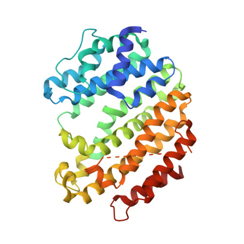
    Zaragozic acids (ZAs) belong to a family of fungal metabolites with nanomolar inhibitory activity toward squalene synthase (SQS). The enzyme catalyzes the committed step of sterol synthesis and has attracted attention as a potential target for antilipogenic and antiinfective therapies. Here, we have determined the structure of ZA-A complexed with human SQS. ZA-A binding induces a local conformational change in the substrate binding site, and its C-6 acyl group also extends over to the cofactor binding cavity. In addition, ZA-A effectively inhibits a homologous bacterial enzyme, dehydrosqualene synthase (CrtM), which synthesizes the precursor of staphyloxanthin in Staphylococcus aureus to cope with oxidative stress. Size reduction at Tyr(248) in CrtM further increases the ZA-A binding affinity, and it reveals a similar overall inhibitor binding mode to that of human SQS/ZA-A except for the C-6 acyl group. These structures pave the way for further improving selectivity and development of a new generation of anticholesterolemic and antimicrobial inhibitors.


  • Organizational Affiliation
    • Institute of Biological Chemistry, Academia Sinica, Taipei 115, Taiwan.

Macromolecule Content 

  • Total Structure Weight: 236.69 kDa 
  • Atom Count: 16,888 
  • Modeled Residue Count: 1,973 
  • Deposited Residue Count: 2,058 
  • Unique protein chains: 1

Macromolecules

Find similar proteins by:|  3D Structure
Entity ID: 1
MoleculeChains  Sequence LengthOrganismDetailsImage
Squalene synthase
A, B, C, D, E
A, B, C, D, E, F
343Homo sapiensMutation(s): 0 
Gene Names: FDFT1
EC: 2.5.1.21
UniProt & NIH Common Fund Data Resources
Find proteins for P37268 (Homo sapiens)
Explore P37268 
Go to UniProtKB:  P37268
PHAROS:  P37268
GTEx:  ENSG00000079459 
Entity Groups
Sequence Clusters30% Identity50% Identity70% Identity90% Identity95% Identity100% Identity
UniProt GroupP37268
Sequence Annotations
Expand
Reference Sequence

Experimental Data & Validation

Experimental Data

  • Method: X-RAY DIFFRACTION
  • Resolution: 2.05 Å
  • R-Value Free:  0.227 (Depositor), 0.228 (DCC) 
  • R-Value Work:  0.169 (Depositor), 0.170 (DCC) 
  • R-Value Observed: 0.172 (Depositor) 
Space Group: P 1 21 1
Unit Cell:
Length ( Å )Angle ( ˚ )
a = 85.61α = 90
b = 153.86β = 91.68
c = 91.516γ = 90
Software Package:
Software NamePurpose
HKL-2000data collection
MOLREPphasing
REFMACrefinement
HKL-2000data reduction
HKL-2000data scaling

Structure Validation

View Full Validation Report


Entry History 

Deposition Data

Revision History  (Full details and data files)

  • Version 1.0: 2012-04-11
    Type: Initial release
  • Version 1.1: 2013-07-24
    Changes: Database references
  • Version 1.2: 2023-11-08
    Changes: Data collection, Database references, Refinement description