3VI2 | pdb_00003vi2

Crystal Structure Analysis of Plasmodium falciparum OMP Decarboxylase in complex with inhibitor HMOA


Experimental Data Snapshot

  • Method: X-RAY DIFFRACTION
  • Resolution: 2.10 Å
  • R-Value Free: 
    0.284 (Depositor), 0.279 (DCC) 
  • R-Value Work: 
    0.213 (Depositor), 0.213 (DCC) 
  • R-Value Observed: 
    0.217 (Depositor) 

wwPDB Validation 3D Report Full Report

Validation slider image for 3VI2

Ligand Structure Quality Assessment 


This is version 1.2 of the entry. See complete history

Literature

The in silico screening and X-ray structure analysis of the inhibitor complex of Plasmodium falciparum orotidine 5'-monophosphate decarboxylase

Takashima, Y.Mizohata, E.Krungkrai, S.R.Fukunishi, Y.Kinoshita, T.Sakata, T.Matsumura, H.Krungkrai, J.Horii, T.Inoue, T.

(2012) J Biochem 152: 133-138

  • DOI: https://doi.org/10.1093/jb/mvs070
  • Primary Citation Related Structures: 
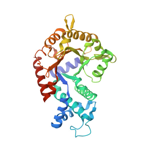
    3VI2

  • PubMed Abstract: 

    Orotidine 5'-monophosphate decarboxylase from Plasmodium falciparum (PfOMPDC) catalyses the final step in the de novo synthesis of uridine 5'-monophosphate (UMP) from orotidine 5'-monophosphate (OMP). A defective PfOMPDC enzyme is lethal to the parasite. Novel in silico screening methods were performed to select 14 inhibitors against PfOMPDC, with a high hit rate of 9%. X-ray structure analysis of PfOMPDC in complex with one of the inhibitors, 4-(2-hydroxy-4-methoxyphenyl)-4-oxobutanoic acid, was carried out to at 2.1 Å resolution. The crystal structure revealed that the inhibitor molecule occupied a part of the active site that overlaps with the phosphate-binding region in the OMP- or UMP-bound complexes. Space occupied by the pyrimidine and ribose rings of OMP or UMP was not occupied by this inhibitor. The carboxyl group of the inhibitor caused a dramatic movement of the L1 and L2 loops that play a role in the recognition of the substrate and product molecules. Combining part of the inhibitor molecule with moieties of the pyrimidine and ribose rings of OMP and UMP represents a suitable avenue for further development of anti-malarial drugs.


  • Organizational Affiliation
    • Department of Applied Chemistry, Graduate School of Engineering, Osaka University, 2-1 Yamadaoka, Suita, Osaka 565-0871, Japan.

Macromolecule Content 

  • Total Structure Weight: 76.25 kDa 
  • Atom Count: 5,385 
  • Modeled Residue Count: 636 
  • Deposited Residue Count: 646 
  • Unique protein chains: 1

Macromolecules

Find similar proteins by:|  3D Structure
Entity ID: 1
MoleculeChains  Sequence LengthOrganismDetailsImage
Orotidine 5'-phosphate decarboxylase
A, B
323Plasmodium falciparumMutation(s): 0 
Gene Names: ompdc
EC: 4.1.1.23
UniProt
Find proteins for Q8T6J6 (Plasmodium falciparum)
Explore Q8T6J6 
Go to UniProtKB:  Q8T6J6
Entity Groups
Sequence Clusters30% Identity50% Identity70% Identity90% Identity95% Identity100% Identity
UniProt GroupQ8T6J6
Sequence Annotations
Expand
Reference Sequence

Experimental Data & Validation

Experimental Data

  • Method: X-RAY DIFFRACTION
  • Resolution: 2.10 Å
  • R-Value Free:  0.284 (Depositor), 0.279 (DCC) 
  • R-Value Work:  0.213 (Depositor), 0.213 (DCC) 
  • R-Value Observed: 0.217 (Depositor) 
Space Group: H 3
Unit Cell:
Length ( Å )Angle ( ˚ )
a = 201.97α = 90
b = 201.97β = 90
c = 44.28γ = 120
Software Package:
Software NamePurpose
HKL-2000data collection
MOLREPphasing
REFMACrefinement
HKL-2000data reduction
HKL-2000data scaling

Structure Validation

View Full Validation Report


Ligand Structure Quality Assessment 


Entry History 

Deposition Data

Revision History  (Full details and data files)

  • Version 1.0: 2012-08-01
    Type: Initial release
  • Version 1.1: 2013-07-17
    Changes: Database references
  • Version 1.2: 2024-03-20
    Changes: Data collection, Database references, Derived calculations