3VHI | pdb_00003vhi

Crystal structure of monoZ-biotin-avidin complex


Experimental Data Snapshot

  • Method: X-RAY DIFFRACTION
  • Resolution: 1.76 Å
  • R-Value Free: 
    0.226 (Depositor), 0.224 (DCC) 
  • R-Value Work: 
    0.184 (Depositor), 0.184 (DCC) 
  • R-Value Observed: 
    0.187 (Depositor) 

Starting Model: experimental
View more details

wwPDB Validation 3D Report Full Report

Validation slider image for 3VHI

Ligand Structure Quality Assessment 


This is version 1.4 of the entry. See complete history

Literature

Rational development of caged-biotin protein-labeling agents and some applications in live cells

Terai, T.Maki, E.Sugiyama, S.Takahashi, Y.Matsumura, H.Mori, Y.Nagano, T.

(2011) Chem Biol 18: 1261-1272

  • DOI: https://doi.org/10.1016/j.chembiol.2011.09.007
  • Primary Citation Related Structures: 
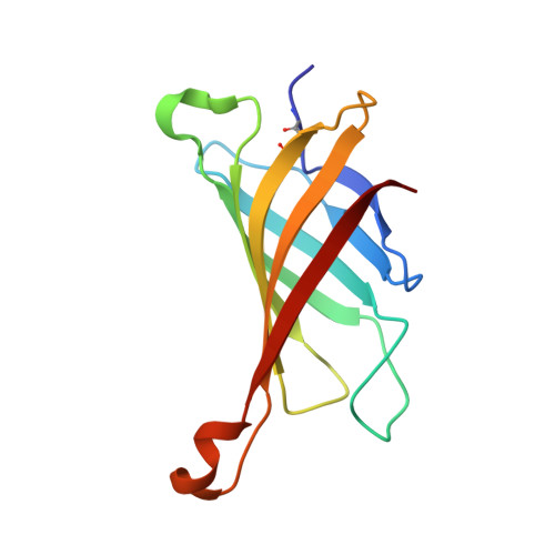
    3VGW, 3VHH, 3VHI, 3VHM

  • PubMed Abstract: 

    Biotin-(strept)avidin complex is widely used in biotechnology because of its extremely high binding constant, but there is no report describing spatiotemporally controlled formation of the complex in live cells. Here, based on X-ray crystal structure analysis and calorimetric data, we designed and synthesized photoreleasable biotins, which show greatly reduced affinity for (strept)avidin, but recover native affinity after UV irradiation. For application at the cell surface, we introduced an amine-reactive moiety into these "caged" biotin molecules. Specific fluorescence imaging of live cells that had been labeled with these agents and then UV-irradiated, was accomplished by addition of streptavidin conjugated with a fluorophore. We also demonstrated the applicability of these compounds for UV-irradiated-cell-specific drug delivery by using caged-biotin-labeled cells, a prodrug, and streptavidin conjugated with a prodrug-activating enzyme.


  • Organizational Affiliation
    • Graduate School of Pharmaceutical Sciences, The University of Tokyo, 7-3-1 Hongo, Bunkyo-ku, Tokyo 113-0033, Japan.

Macromolecule Content 

  • Total Structure Weight: 57.56 kDa 
  • Atom Count: 4,178 
  • Modeled Residue Count: 486 
  • Deposited Residue Count: 488 
  • Unique protein chains: 1

Macromolecules

Find similar proteins by:|  3D Structure
Entity ID: 1
MoleculeChains  Sequence LengthOrganismDetailsImage
Avidin
A, B, C, D
122Gallus gallusMutation(s): 0 
UniProt
Find proteins for P02701 (Gallus gallus)
Explore P02701 
Go to UniProtKB:  P02701
Entity Groups
Sequence Clusters30% Identity50% Identity70% Identity90% Identity95% Identity100% Identity
UniProt GroupP02701
Glycosylation
Glycosylation Sites: 1
Sequence Annotations
Expand
Reference Sequence

Small Molecules

Ligands 3 Unique
IDChains Name / Formula / InChI Key2D Diagram3D Interactions
VHI

Query on VHI


Download:Ideal Coordinates CCD File
E [auth A],
I [auth B],
M [auth C],
P [auth D]
5-{(3aS,4S,6aR)-1-[(benzyloxy)carbonyl]-2-oxohexahydro-1H-thieno[3,4-d]imidazol-4-yl}pentanoic acid
C18 H22 N2 O5 S
BWNGWJMPVBQLHD-DZKIICNBSA-N
NAG

Query on NAG


Download:Ideal Coordinates CCD File
F [auth A],
J [auth B],
N [auth C],
Q [auth D]
2-acetamido-2-deoxy-beta-D-glucopyranose
C8 H15 N O6
OVRNDRQMDRJTHS-FMDGEEDCSA-N
SO4

Query on SO4


Download:Ideal Coordinates CCD File
G [auth A]
H [auth A]
K [auth B]
L [auth B]
O [auth C]
G [auth A],
H [auth A],
K [auth B],
L [auth B],
O [auth C],
R [auth D]
SULFATE ION
O4 S
QAOWNCQODCNURD-UHFFFAOYSA-L

Experimental Data & Validation

Experimental Data

  • Method: X-RAY DIFFRACTION
  • Resolution: 1.76 Å
  • R-Value Free:  0.226 (Depositor), 0.224 (DCC) 
  • R-Value Work:  0.184 (Depositor), 0.184 (DCC) 
  • R-Value Observed: 0.187 (Depositor) 
Space Group: C 1 2 1
Unit Cell:
Length ( Å )Angle ( ˚ )
a = 130.781α = 90
b = 73.455β = 120.05
c = 81.251γ = 90
Software Package:
Software NamePurpose
DENZOdata reduction
SCALEPACKdata scaling
REFMACrefinement
PDB_EXTRACTdata extraction
HKL-2000data reduction
MOLREPphasing

Structure Validation

View Full Validation Report


Ligand Structure Quality Assessment 


Entry History 

Deposition Data

Revision History  (Full details and data files)

  • Version 1.0: 2011-12-28
    Type: Initial release
  • Version 1.1: 2017-11-22
    Changes: Refinement description
  • Version 1.2: 2020-07-29
    Type: Remediation
    Reason: Carbohydrate remediation
    Changes: Data collection, Derived calculations, Structure summary
  • Version 1.3: 2023-11-08
    Changes: Data collection, Database references, Refinement description, Structure summary
  • Version 1.4: 2024-10-16
    Changes: Structure summary