3VHF | pdb_00003vhf

plant thaumatin I at pH 8.0


Experimental Data Snapshot

  • Method: X-RAY DIFFRACTION
  • Resolution: 1.39 Å
  • R-Value Free: 
    0.168 (Depositor), 0.168 (DCC) 
  • R-Value Work: 
    0.148 (Depositor), 0.147 (DCC) 
  • R-Value Observed: 
    0.149 (Depositor) 

Starting Model: experimental
View more details

wwPDB Validation 3D Report Full Report

Validation slider image for 3VHF

This is version 1.2 of the entry. See complete history

Literature

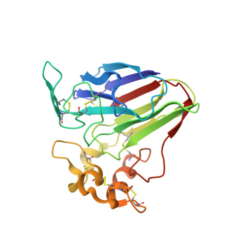
Atomic structure of the sweet-tasting protein thaumatin I at pH 8.0 reveals the large disulfide-rich region in domain II to be sensitive to a pH change

Masuda, T.Ohta, K.Mikami, B.Kitabatake, N.Tani, F.

(2012) Biochem Biophys Res Commun 419: 72-76

  • DOI: https://doi.org/10.1016/j.bbrc.2012.01.129
  • Primary Citation Related Structures: 
    3VHF, 3VJQ

  • PubMed Abstract: 

    Thaumatin, an intensely sweet-tasting plant protein, elicits a sweet taste at 50 nM. Although the sweetness remains when thaumatin is heated at 80 °C for 4h under acid conditions, it rapidly declines when heating at a pH above 6.5. To clarify the structural difference at high pH, the atomic structure of a recombinant thaumatin I at pH 8.0 was determined at a resolution of 1.0Å. Comparison to the crystal structure of thaumatin at pH 7.3 and 7.0 revealed the root-mean square deviation value of a Cα atom to be substantially greater in the large disulfide-rich region of domain II, especially residues 154-164, suggesting that a loop region in domain II to be affected by solvent conditions. Furthermore, B-factors of Lys137, Lys163, and Lys187 were significantly affected by pH change, suggesting that a striking increase in the mobility of these lysine residues, which could facilitate a reaction with a free sulfhydryl residue produced via the β-elimination of disulfide bonds by heating at a pH above 7.0. The increase in mobility of lysine residues as well as a loop region in domain II might play an important role in the heat-induced aggregation of thaumatin above pH 7.0.


  • Organizational Affiliation
    • Division of Food Science and Biotechnology, Graduate School of Agriculture, Kyoto University, Gokasho, Uji, Kyoto 611-0011, Japan. t2masuda@kais.kyoto-u.ac.jp

Macromolecule Content 

  • Total Structure Weight: 22.41 kDa 
  • Atom Count: 2,104 
  • Modeled Residue Count: 207 
  • Deposited Residue Count: 207 
  • Unique protein chains: 1

Macromolecules

Find similar proteins by:|  3D Structure
Entity ID: 1
MoleculeChains  Sequence LengthOrganismDetailsImage
Thaumatin I207Thaumatococcus danielliiMutation(s): 0 
UniProt
Find proteins for P02883 (Thaumatococcus daniellii)
Explore P02883 
Go to UniProtKB:  P02883
Entity Groups
Sequence Clusters30% Identity50% Identity70% Identity90% Identity95% Identity100% Identity
UniProt GroupP02883
Sequence Annotations
Expand
Reference Sequence

Experimental Data & Validation

Experimental Data

  • Method: X-RAY DIFFRACTION
  • Resolution: 1.39 Å
  • R-Value Free:  0.168 (Depositor), 0.168 (DCC) 
  • R-Value Work:  0.148 (Depositor), 0.147 (DCC) 
  • R-Value Observed: 0.149 (Depositor) 
Space Group: P 21 21 21
Unit Cell:
Length ( Å )Angle ( ˚ )
a = 43.688α = 90
b = 63.653β = 90
c = 71.743γ = 90
Software Package:
Software NamePurpose
REFMACrefinement
HKL-2000data reduction
HKL-2000data scaling

Structure Validation

View Full Validation Report


Entry History 

Deposition Data

Revision History  (Full details and data files)

  • Version 1.0: 2012-05-16
    Type: Initial release
  • Version 1.1: 2023-11-08
    Changes: Data collection, Database references, Derived calculations, Refinement description
  • Version 1.2: 2024-11-06
    Changes: Structure summary