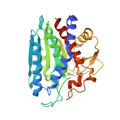
3VH9

Crystal structure of Aeromonas proteolytica aminopeptidase complexed with 8-quinolinol


Experimental Data Snapshot

  • Method: X-RAY DIFFRACTION
  • Resolution: 1.29 Å
  • R-Value Free: 0.153 
  • R-Value Work: 0.131 
  • R-Value Observed: 0.132 

Starting Model: experimental
View more details

wwPDB Validation   3D Report Full Report


This is version 1.1 of the entry. See complete history


Literature

Potent inhibition of dinuclear zinc(II) peptidase, an aminopeptidase from Aeromonas proteolytica, by 8-quinolinol derivatives: inhibitor design based on Zn(2+) fluorophores, kinetic, and X-ray crystallographic study.

Hanaya, K.Suetsugu, M.Saijo, S.Yamato, I.Aoki, S.

(2012) J Biol Inorg Chem 17: 517-529

  • DOI: https://doi.org/10.1007/s00775-012-0873-4
  • Primary Citation of Related Structures:  
    3VH9

  • PubMed Abstract: 

    The selective inhibition of an aminopeptidase from Aeromonas proteolytica (AAP), a dinuclear Zn(2+) hydrolase, by 8-quinolinol (8-hydroxyquinoline, 8-HQ) derivatives is reported. We previously reported on the preparation of 8-HQ-pendant cyclens as Zn(2+) fluorophores (cyclen is 1,4,7,10-tetraazacyclododecane), in which the nitrogen and phenolate of the 8-HQ units (as well as the four nitrogens of cyclen) bind to Zn(2+) in a bidentate manner to form very stable Zn(2+) complexes at neutral pH (K (d) = 8-50 fM at pH 7.4). On the basis of this finding, it was hypothesized that 8-HQ derivatives have the potential to function as specific inhibitors of Zn(2+) enzymes, especially dinuclear Zn(2+) hydrolases. Assays of 8-HQ derivatives as inhibitors were performed against commercially available dinuclear Zn(2+) enzymes such as AAP and alkaline phosphatase. 8-HQ and the 5-substituted 8-HQ derivatives were found to be competitive inhibitors of AAP with inhibition constants of 0.16-29 μM at pH 8.0. The nitrogen at the 1-position and the hydroxide at the 8-position of 8-HQ were found to be essential for the inhibition of AAP. Fluorescence titrations of these drugs with AAP and an X-ray crystal structure analysis of an AAP-8-HQ complex (1.3-Å resolution) confirmed that 8-HQ binds to AAP in the "Pyr-out" mode, in which the hydroxide anion of 8-HQ bridges two Zn(2+) ions (Zn1 and Zn2) in the active site of AAP and the nitrogen atom of 8-HQ coordinates to Zn1 (Protein Data Bank code 3VH9).


  • Organizational Affiliation

    Faculty of Pharmaceutical Sciences, Tokyo University of Science, 2641 Yamazaki, Noda 278-8510, Japan.


Macromolecules
Find similar proteins by:  (by identity cutoff)  |  3D Structure
Entity ID: 1
MoleculeChains Sequence LengthOrganismDetailsImage
Bacterial leucyl aminopeptidase299Vibrio proteolyticusMutation(s): 0 
EC: 3.4.11.10
UniProt
Find proteins for Q01693 (Vibrio proteolyticus)
Explore Q01693 
Go to UniProtKB:  Q01693
Entity Groups  
Sequence Clusters30% Identity50% Identity70% Identity90% Identity95% Identity100% Identity
UniProt GroupQ01693
Sequence Annotations
Expand
  • Reference Sequence
Small Molecules
Ligands 6 Unique
IDChains Name / Formula / InChI Key2D Diagram3D Interactions
HQY
Query on HQY

Download Ideal Coordinates CCD File 
D [auth A]quinolin-8-ol
C9 H7 N O
MCJGNVYPOGVAJF-UHFFFAOYSA-N
GOL
Query on GOL

Download Ideal Coordinates CCD File 
Y [auth A]GLYCEROL
C3 H8 O3
PEDCQBHIVMGVHV-UHFFFAOYSA-N
ZN
Query on ZN

Download Ideal Coordinates CCD File 
B [auth A],
C [auth A]
ZINC ION
Zn
PTFCDOFLOPIGGS-UHFFFAOYSA-N
SCN
Query on SCN

Download Ideal Coordinates CCD File 
W [auth A],
X [auth A]
THIOCYANATE ION
C N S
ZMZDMBWJUHKJPS-UHFFFAOYSA-M
CL
Query on CL

Download Ideal Coordinates CCD File 
N [auth A]
O [auth A]
P [auth A]
Q [auth A]
R [auth A]
N [auth A],
O [auth A],
P [auth A],
Q [auth A],
R [auth A],
S [auth A],
T [auth A],
U [auth A],
V [auth A]
CHLORIDE ION
Cl
VEXZGXHMUGYJMC-UHFFFAOYSA-M
NA
Query on NA

Download Ideal Coordinates CCD File 
E [auth A]
F [auth A]
G [auth A]
H [auth A]
I [auth A]
E [auth A],
F [auth A],
G [auth A],
H [auth A],
I [auth A],
J [auth A],
K [auth A],
L [auth A],
M [auth A]
SODIUM ION
Na
FKNQFGJONOIPTF-UHFFFAOYSA-N
Binding Affinity Annotations 
IDSourceBinding Affinity
HQY PDBBind:  3VH9 Ki: 580 (nM) from 1 assay(s)
Experimental Data & Validation

Experimental Data

  • Method: X-RAY DIFFRACTION
  • Resolution: 1.29 Å
  • R-Value Free: 0.153 
  • R-Value Work: 0.131 
  • R-Value Observed: 0.132 
  • Space Group: P 61 2 2
Unit Cell:
Length ( Å )Angle ( ˚ )
a = 108.463α = 90
b = 108.463β = 90
c = 90.737γ = 120
Software Package:
Software NamePurpose
HKL-2000data collection
PHASERphasing
PHENIXrefinement
REFMACrefinement
HKL-2000data reduction
HKL-2000data scaling

Structure Validation

View Full Validation Report


Entry History 

Deposition Data

Revision History  (Full details and data files)

  • Version 1.0: 2012-05-02
    Type: Initial release
  • Version 1.1: 2023-11-08
    Changes: Data collection, Database references, Derived calculations, Refinement description