3VH4

Crystal structure of Atg7CTD-Atg8-MgATP complex


Experimental Data Snapshot

  • Method: X-RAY DIFFRACTION
  • Resolution: 2.65 Å
  • R-Value Free: 0.267 
  • R-Value Work: 0.235 
  • R-Value Observed: 0.235 

Starting Model: experimental
View more details

wwPDB Validation   3D Report Full Report


Ligand Structure Quality Assessment 


This is version 1.3 of the entry. See complete history


Literature

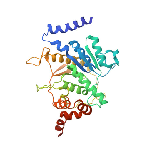
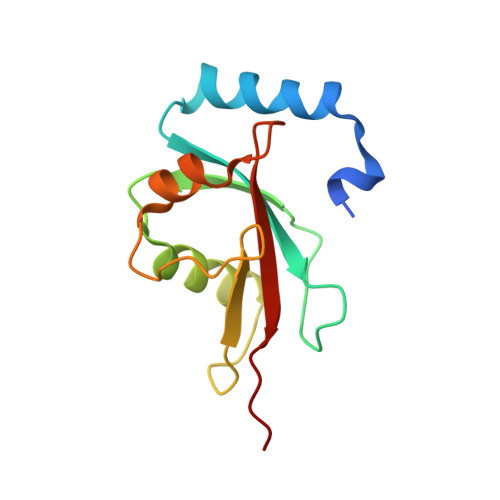
Structural basis of Atg8 activation by a homodimeric E1, Atg7.

Noda, N.N.Satoo, K.Fujioka, Y.Kumeta, H.Ogura, K.Nakatogawa, H.Ohsumi, Y.Inagaki, F.

(2011) Mol Cell 44: 462-475

  • DOI: https://doi.org/10.1016/j.molcel.2011.08.035
  • Primary Citation of Related Structures:  
    2LI5, 3VH1, 3VH2, 3VH3, 3VH4

  • PubMed Abstract: 

    E1 enzymes activate ubiquitin-like proteins and transfer them to cognate E2 enzymes. Atg7, a noncanonical E1, activates two ubiquitin-like proteins, Atg8 and Atg12, and plays a crucial role in autophagy. Here, we report crystal structures of full-length Atg7 and its C-terminal domain bound to Atg8 and MgATP, as well as a solution structure of Atg8 bound to the extreme C-terminal domain (ECTD) of Atg7. The unique N-terminal domain (NTD) of Atg7 is responsible for Atg3 (E2) binding, whereas its C-terminal domain is comprised of a homodimeric adenylation domain (AD) and ECTD. The structural and biochemical data demonstrate that Atg8 is initially recognized by the C-terminal tail of ECTD and is then transferred to an AD, where the Atg8 C terminus is attacked by the catalytic cysteine to form a thioester bond. Atg8 is then transferred via a trans mechanism to the Atg3 bound to the NTD of the opposite protomer within a dimer.


  • Organizational Affiliation

    Institute of Microbial Chemistry, Tokyo, Tokyo 141-0021, Japan. nn@bikaken.or.jp


Macromolecules
Find similar proteins by:  (by identity cutoff)  |  3D Structure
Entity ID: 1
MoleculeChains Sequence LengthOrganismDetailsImage
Ubiquitin-like modifier-activating enzyme ATG7340Saccharomyces cerevisiae S288CMutation(s): 0 
Gene Names: ATG7APG7CVT2YHR171W
UniProt
Find proteins for P38862 (Saccharomyces cerevisiae (strain ATCC 204508 / S288c))
Explore P38862 
Go to UniProtKB:  P38862
Entity Groups  
Sequence Clusters30% Identity50% Identity70% Identity90% Identity95% Identity100% Identity
UniProt GroupP38862
Sequence Annotations
Expand
  • Reference Sequence
Find similar proteins by:  (by identity cutoff)  |  3D Structure
Entity ID: 2
MoleculeChains Sequence LengthOrganismDetailsImage
Autophagy-related protein 8119Saccharomyces cerevisiae S288CMutation(s): 1 
Gene Names: ATG8APG8AUT7CVT5YBL078CYBL0732
UniProt
Find proteins for P38182 (Saccharomyces cerevisiae (strain ATCC 204508 / S288c))
Explore P38182 
Go to UniProtKB:  P38182
Entity Groups  
Sequence Clusters30% Identity50% Identity70% Identity90% Identity95% Identity100% Identity
UniProt GroupP38182
Sequence Annotations
Expand
  • Reference Sequence
Experimental Data & Validation

Experimental Data

  • Method: X-RAY DIFFRACTION
  • Resolution: 2.65 Å
  • R-Value Free: 0.267 
  • R-Value Work: 0.235 
  • R-Value Observed: 0.235 
  • Space Group: P 43 21 2
Unit Cell:
Length ( Å )Angle ( ˚ )
a = 69.282α = 90
b = 69.282β = 90
c = 219.605γ = 90
Software Package:
Software NamePurpose
HKL-2000data collection
CNSrefinement
HKL-2000data reduction
HKL-2000data scaling
CNSphasing

Structure Validation

View Full Validation Report


Ligand Structure Quality Assessment 


Entry History 

Deposition Data

Revision History  (Full details and data files)

  • Version 1.0: 2011-09-21
    Type: Initial release
  • Version 1.1: 2011-11-16
    Changes: Database references
  • Version 1.2: 2012-03-28
    Changes: Database references
  • Version 1.3: 2023-11-08
    Changes: Data collection, Database references, Derived calculations, Refinement description