3VFK

The structure of monodechloro-teicoplanin in complex with its ligand, using ubiquitin as a ligand carrier


Experimental Data Snapshot

  • Method: X-RAY DIFFRACTION
  • Resolution: 2.80 Å
  • R-Value Free: 0.306 
  • R-Value Work: 0.278 
  • R-Value Observed: 0.279 

Starting Model: experimental
View more details

wwPDB Validation   3D Report Full Report


Ligand Structure Quality Assessment 


This is version 3.1 of the entry. See complete history


Literature

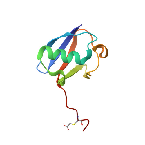
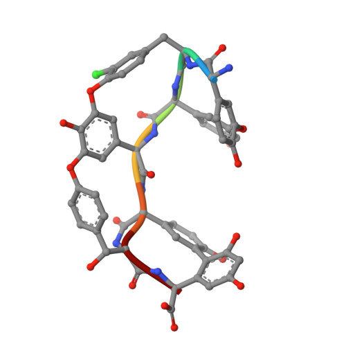
Structure of the complex between teicoplanin and a bacterial cell-wall peptide: use of a carrier-protein approach.

Economou, N.J.Zentner, I.J.Lazo, E.Jakoncic, J.Stojanoff, V.Weeks, S.D.Grasty, K.C.Cocklin, S.Loll, P.J.

(2013) Acta Crystallogr D Biol Crystallogr 69: 520-533

  • DOI: https://doi.org/10.1107/S0907444912050469
  • Primary Citation of Related Structures:  
    3VFJ, 3VFK

  • PubMed Abstract: 

    Multidrug-resistant bacterial infections are commonly treated with glycopeptide antibiotics such as teicoplanin. This drug inhibits bacterial cell-wall biosynthesis by binding and sequestering a cell-wall precursor: a D-alanine-containing peptide. A carrier-protein strategy was used to crystallize the complex of teicoplanin and its target peptide by fusing the cell-wall peptide to either MBP or ubiquitin via native chemical ligation and subsequently crystallizing the protein-peptide-antibiotic complex. The 2.05 Å resolution MBP-peptide-teicoplanin structure shows that teicoplanin recognizes its ligand through a combination of five hydrogen bonds and multiple van der Waals interactions. Comparison of this teicoplanin structure with that of unliganded teicoplanin reveals a flexibility in the antibiotic peptide backbone that has significant implications for ligand recognition. Diffraction experiments revealed an X-ray-induced dechlorination of the sixth amino acid of the antibiotic; it is shown that teicoplanin is significantly more radiation-sensitive than other similar antibiotics and that ligand binding increases radiosensitivity. Insights derived from this new teicoplanin structure may contribute to the development of next-generation antibacterials designed to overcome bacterial resistance.


  • Organizational Affiliation

    Department of Biochemistry and Molecular Biology, Drexel University College of Medicine, Philadelphia, PA 19102, USA.


Macromolecules
Find similar proteins by:  (by identity cutoff)  |  3D Structure
Entity ID: 1
MoleculeChains Sequence LengthOrganismDetailsImage
ubiquitin, C-terminal fused by Cys-Lys-D-Ala-D-Ala79Homo sapiensMutation(s): 0 
Gene Names: UBC
UniProt & NIH Common Fund Data Resources
Find proteins for P0CG48 (Homo sapiens)
Explore P0CG48 
Go to UniProtKB:  P0CG48
GTEx:  ENSG00000150991 
Entity Groups  
Sequence Clusters30% Identity50% Identity70% Identity90% Identity95% Identity100% Identity
UniProt GroupP0CG48
Sequence Annotations
Expand
  • Reference Sequence

Find similar proteins by:  Sequence   |   3D Structure  

Entity ID: 2
MoleculeChains Sequence LengthOrganismDetailsImage
MonodeChloro- Teicoplanin A2-2B [auth G]7Actinoplanes teichomyceticusMutation(s): 0 
Sequence Annotations
Expand
  • Reference Sequence
Small Molecules
Modified Residues  3 Unique
IDChains TypeFormula2D DiagramParent
CCS
Query on CCS
A
L-PEPTIDE LINKINGC5 H9 N O4 SCYS
3MY
Query on 3MY
B [auth G]D-PEPTIDE LINKINGC9 H10 Cl N O3TYR
OMX
Query on OMX
B [auth G]L-PEPTIDE LINKINGC9 H11 N O4TYR
Biologically Interesting Molecules (External Reference) 1 Unique
Experimental Data & Validation

Experimental Data

  • Method: X-RAY DIFFRACTION
  • Resolution: 2.80 Å
  • R-Value Free: 0.306 
  • R-Value Work: 0.278 
  • R-Value Observed: 0.279 
  • Space Group: C 1 2 1
Unit Cell:
Length ( Å )Angle ( ˚ )
a = 88.01α = 90
b = 25.18β = 109.18
c = 38.71γ = 90
Software Package:
Software NamePurpose
MOLREPphasing
PHENIXrefinement
XDSdata reduction
XSCALEdata scaling

Structure Validation

View Full Validation Report


Ligand Structure Quality Assessment 


Entry History 

Deposition Data

Revision History  (Full details and data files)

  • Version 1.0: 2013-01-09
    Type: Initial release
  • Version 1.1: 2013-03-27
    Changes: Structure summary
  • Version 1.2: 2013-04-03
    Changes: Derived calculations
  • Version 1.3: 2013-06-19
    Changes: Database references
  • Version 2.0: 2020-07-29
    Type: Remediation
    Reason: Carbohydrate remediation
    Changes: Data collection, Derived calculations, Polymer sequence, Structure summary
  • Version 2.1: 2023-09-13
    Changes: Data collection, Database references, Refinement description, Structure summary
  • Version 3.0: 2023-09-27
    Changes: Polymer sequence
  • Version 3.1: 2023-12-06
    Changes: Data collection, Derived calculations