3VEM | pdb_00003vem

Structural basis of transcriptional gene silencing mediated by Arabidopsis MOM1


Experimental Data Snapshot

  • Method: X-RAY DIFFRACTION
  • Resolution: 3.20 Å
  • R-Value Free: 
    0.293 (Depositor), 0.296 (DCC) 
  • R-Value Work: 
    0.258 (Depositor), 0.263 (DCC) 
  • R-Value Observed: 
    0.260 (Depositor) 

wwPDB Validation 3D Report Full Report

Validation slider image for 3VEM

This is version 1.1 of the entry. See complete history

Literature

Structural Basis of Transcriptional Gene Silencing Mediated by Arabidopsis MOM1.

Nishimura, T.Molinard, G.Petty, T.J.Broger, L.Gabus, C.Halazonetis, T.D.Thore, S.Paszkowski, J.

(2012) PLoS Genet 8: e1002484-e1002484

  • DOI: https://doi.org/10.1371/journal.pgen.1002484
  • Primary Citation Related Structures: 
    3VEM

  • PubMed Abstract: 

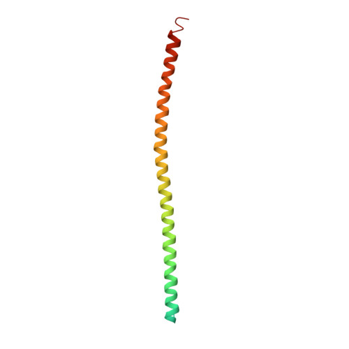
    Shifts between epigenetic states of transcriptional activity are typically correlated with changes in epigenetic marks. However, exceptions to this rule suggest the existence of additional, as yet uncharacterized, layers of epigenetic regulation. MOM1, a protein of 2,001 amino acids that acts as a transcriptional silencer, represents such an exception. Here we define the 82 amino acid domain called CMM2 (Conserved MOM1 Motif 2) as a minimal MOM1 fragment capable of transcriptional regulation. As determined by X-ray crystallography, this motif folds into an unusual hendecad-based coiled-coil. Structure-based mutagenesis followed by transgenic complementation tests in plants demonstrate that CMM2 and its dimerization are effective for transcriptional suppression at chromosomal loci co-regulated by MOM1 and the siRNA pathway but not at loci controlled by MOM1 in an siRNA-independent fashion. These results reveal a surprising separation of epigenetic activities that enable the single, large MOM1 protein to coordinate cooperating mechanisms of epigenetic regulation.


  • Organizational Affiliation
    • Department of Plant Biology, University of Geneva, Geneva, Switzerland.

Macromolecule Content 

  • Total Structure Weight: 53.04 kDa 
  • Atom Count: 2,761 
  • Modeled Residue Count: 327 
  • Deposited Residue Count: 460 
  • Unique protein chains: 1

Macromolecules

Find similar proteins by:|  3D Structure
Entity ID: 1
MoleculeChains  Sequence LengthOrganismDetailsImage
Helicase protein MOM1
A, B, C, D
115Arabidopsis thalianaMutation(s): 0 
Gene Names: MOM1MOMAt1g08060T6D22.14
EC: 3.6.4
UniProt
Find proteins for Q9M658 (Arabidopsis thaliana)
Explore Q9M658 
Go to UniProtKB:  Q9M658
Entity Groups
Sequence Clusters30% Identity50% Identity70% Identity90% Identity95% Identity100% Identity
UniProt GroupQ9M658
Sequence Annotations
Expand
Reference Sequence

Experimental Data & Validation

Experimental Data

  • Method: X-RAY DIFFRACTION
  • Resolution: 3.20 Å
  • R-Value Free:  0.293 (Depositor), 0.296 (DCC) 
  • R-Value Work:  0.258 (Depositor), 0.263 (DCC) 
  • R-Value Observed: 0.260 (Depositor) 
Space Group: P 31 2 1
Unit Cell:
Length ( Å )Angle ( ˚ )
a = 85.64α = 90
b = 85.64β = 90
c = 292.74γ = 120
Software Package:
Software NamePurpose
ADSCdata collection
SHARPphasing
PHENIXrefinement
XDSdata reduction
XSCALEdata scaling

Structure Validation

View Full Validation Report


Entry History 

Deposition Data

Revision History  (Full details and data files)

  • Version 1.0: 2012-03-07
    Type: Initial release
  • Version 1.1: 2024-02-28
    Changes: Data collection, Database references