3VC1 | pdb_00003vc1

Crystal structure of geranyl diphosphate C-methyltransferase from Streptomyces coelicolor A3(2) in complex with Mg2+, geranyl-S-thiolodiphosphate, and S-adenosyl-L-homocysteine


Experimental Data Snapshot

  • Method: X-RAY DIFFRACTION
  • Resolution: 1.82 Å
  • R-Value Free: 
    0.194 (Depositor), 0.191 (DCC) 
  • R-Value Work: 
    0.167 (Depositor), 0.163 (DCC) 
  • R-Value Observed: 
    0.167 (Depositor) 

wwPDB Validation 3D Report Full Report

Validation slider image for 3VC1

Ligand Structure Quality Assessment 


This is version 1.3 of the entry. See complete history

Literature

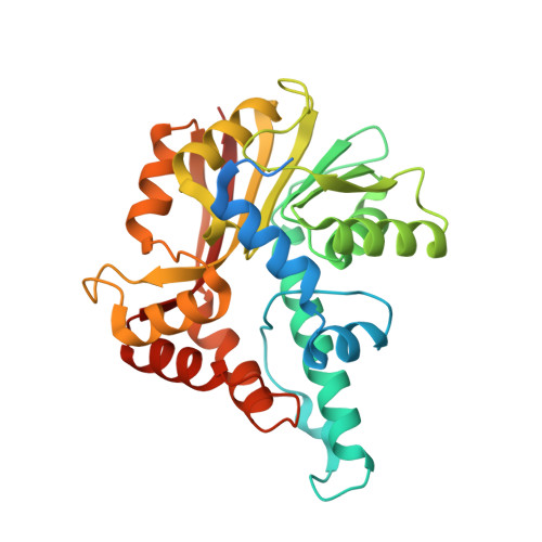
Structure of Geranyl Diphosphate C-Methyltransferase from Streptomyces coelicolor and Implications for the Mechanism of Isoprenoid Modification.

Koksal, M.Chou, W.K.Cane, D.E.Christianson, D.W.

(2012) Biochemistry 51: 3003-3010

  • DOI: https://doi.org/10.1021/bi300109c
  • Primary Citation Related Structures: 
    3VC1, 3VC2

  • PubMed Abstract: 

    Geranyl diphosphate C-methyltransferase (GPPMT) from Streptomyces coelicolor A3(2) is the first methyltransferase discovered that modifies an acyclic isoprenoid diphosphate, geranyl diphosphate (GPP), to yield a noncanonical acyclic allylic diphosphate product, 2-methylgeranyl diphosphate, which serves as the substrate for a subsequent cyclization reaction catalyzed by a terpenoid cyclase, methylisoborneol synthase. Here, we report the crystal structures of GPPMT in complex with GPP or the substrate analogue geranyl S-thiolodiphosphate (GSPP) along with S-adenosyl-L-homocysteine in the cofactor binding site, resulting from in situ demethylation of S-adenosyl-L-methionine, at 2.05 or 1.82 Å resolution, respectively. These structures suggest that both GPP and GSPP can undergo catalytic methylation in crystalline GPPMT, followed by dissociation of the isoprenoid product. S-Adenosyl-L-homocysteine remains bound in the active site, however, and does not exchange with a fresh molecule of cofactor S-adenosyl-L-methionine. These structures provide important clues about the molecular mechanism of the reaction, especially with regard to the face of the 2,3 double bond of GPP that is methylated as well as the stabilization of the resulting carbocation intermediate through cation-π interactions.


  • Organizational Affiliation
    • Roy and Diana Vagelos Laboratories, Department of Chemistry, University of Pennsylvania, 231 South 34th Street, Philadelphia, Pennsylvania 19104-6323, USA.

Macromolecule Content 

  • Total Structure Weight: 430.83 kDa 
  • Atom Count: 29,215 
  • Modeled Residue Count: 3,343 
  • Deposited Residue Count: 3,744 
  • Unique protein chains: 1

Macromolecules

Find similar proteins by:|  3D Structure
Entity ID: 1
MoleculeChains  Sequence LengthOrganismDetailsImage
Geranyl diphosphate 2-C-methyltransferase
A, B, C, D, E
A, B, C, D, E, F, G, H, I, J, K, L
312Streptomyces coelicolor A3(2)Mutation(s): 0 
Gene Names: orf4SCBAC12C8.02SCO7701
EC: 2.1.1 (PDB Primary Data), 2.1.1.255 (UniProt)
UniProt
Find proteins for Q9F1Y5 (Streptomyces coelicolor (strain ATCC BAA-471 / A3(2) / M145))
Explore Q9F1Y5 
Go to UniProtKB:  Q9F1Y5
Entity Groups
Sequence Clusters30% Identity50% Identity70% Identity90% Identity95% Identity100% Identity
UniProt GroupQ9F1Y5
Sequence Annotations
Expand
Reference Sequence

Small Molecules

Ligands 5 Unique
IDChains Name / Formula / InChI Key2D Diagram3D Interactions
SAH

Query on SAH


Download:Ideal Coordinates CCD File
CA [auth D]
CB [auth J]
GA [auth E]
GB [auth K]
JB [auth L]
CA [auth D],
CB [auth J],
GA [auth E],
GB [auth K],
JB [auth L],
MA [auth F],
N [auth A],
QA [auth G],
T [auth B],
UA [auth H],
X [auth C],
YA [auth I]
S-ADENOSYL-L-HOMOCYSTEINE
C14 H20 N6 O5 S
ZJUKTBDSGOFHSH-WFMPWKQPSA-N
GST

Query on GST


Download:Ideal Coordinates CCD File
DA [auth D]
DB [auth J]
HA [auth E]
HB [auth K]
KB [auth L]
DA [auth D],
DB [auth J],
HA [auth E],
HB [auth K],
KB [auth L],
NA [auth F],
O [auth A],
RA [auth G],
U [auth B],
VA [auth H],
Y [auth C],
ZA [auth I]
GERANYL S-THIOLODIPHOSPHATE
C10 H20 O6 P2 S
AKIXWSDUEPPMKM-JXMROGBWSA-N
SO4

Query on SO4


Download:Ideal Coordinates CCD File
IA [auth E]SULFATE ION
O4 S
QAOWNCQODCNURD-UHFFFAOYSA-L
GOL

Query on GOL


Download:Ideal Coordinates CCD File
AA [auth C]
AB [auth I]
EA [auth D]
EB [auth J]
JA [auth E]
AA [auth C],
AB [auth I],
EA [auth D],
EB [auth J],
JA [auth E],
KA [auth E],
LB [auth L],
OA [auth F],
P [auth A],
Q [auth A],
R [auth A],
SA [auth G],
V [auth B],
WA [auth H],
Z [auth C]
GLYCEROL
C3 H8 O3
PEDCQBHIVMGVHV-UHFFFAOYSA-N
MG

Query on MG


Download:Ideal Coordinates CCD File
BA [auth D]
BB [auth J]
FA [auth E]
FB [auth K]
IB [auth L]
BA [auth D],
BB [auth J],
FA [auth E],
FB [auth K],
IB [auth L],
LA [auth F],
M [auth A],
PA [auth G],
S [auth B],
TA [auth H],
W [auth C],
XA [auth I]
MAGNESIUM ION
Mg
JLVVSXFLKOJNIY-UHFFFAOYSA-N

Experimental Data & Validation

Experimental Data

  • Method: X-RAY DIFFRACTION
  • Resolution: 1.82 Å
  • R-Value Free:  0.194 (Depositor), 0.191 (DCC) 
  • R-Value Work:  0.167 (Depositor), 0.163 (DCC) 
  • R-Value Observed: 0.167 (Depositor) 
Space Group: P 1 21 1
Unit Cell:
Length ( Å )Angle ( ˚ )
a = 97.029α = 90
b = 102.903β = 99.59
c = 203.582γ = 90
Software Package:
Software NamePurpose
CBASSdata collection
HKL2Mapmodel building
PHENIXrefinement
HKL-2000data reduction
HKL-2000data scaling
HKL2Mapphasing

Structure Validation

View Full Validation Report


Ligand Structure Quality Assessment 


Entry History 

Deposition Data

Revision History  (Full details and data files)

  • Version 1.0: 2012-04-11
    Type: Initial release
  • Version 1.1: 2012-04-25
    Changes: Database references
  • Version 1.2: 2017-11-08
    Changes: Refinement description
  • Version 1.3: 2024-02-28
    Changes: Data collection, Database references, Derived calculations