3VBQ | pdb_00003vbq

Exploitation of hydrogen bonding constraints and flat hydrophobic energy landscapes in Pim-1 kinase needle screening and inhibitor design


Experimental Data Snapshot

  • Method: X-RAY DIFFRACTION
  • Resolution: 1.85 Å
  • R-Value Free: 
    0.206 (Depositor), 0.205 (DCC) 
  • R-Value Work: 
    0.176 (Depositor), 0.177 (DCC) 
  • R-Value Observed: 
    0.178 (Depositor) 

wwPDB Validation 3D Report Full Report

Validation slider image for 3VBQ

Ligand Structure Quality Assessment 


This is version 2.1 of the entry. See complete history

Literature

Implications of promiscuous Pim-1 kinase fragment inhibitor hydrophobic interactions for fragment-based drug design.

Good, A.C.Liu, J.Hirth, B.Asmussen, G.Xiang, Y.Biemann, H.P.Bishop, K.A.Fremgen, T.Fitzgerald, M.Gladysheva, T.Jain, A.Jancsics, K.Metz, M.Papoulis, A.Skerlj, R.Stepp, J.D.Wei, R.R.

(2012) J Med Chem 55: 2641-2648

  • DOI: https://doi.org/10.1021/jm2014698
  • Primary Citation Related Structures: 
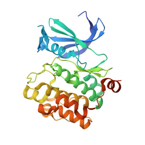
    3VBQ, 3VBT, 3VBV, 3VBW, 3VBX, 3VBY, 3VC4

  • PubMed Abstract: 

    We have studied the subtleties of fragment docking and binding using data generated in a Pim-1 kinase inhibitor program. Crystallographic and docking data analyses have been undertaken using inhibitor complexes derived from an in-house surface plasmon resonance (SPR) fragment screen, a virtual needle screen, and a de novo designed fragment inhibitor hybrid. These investigations highlight that fragments that do not fill their binding pocket can exhibit promiscuous hydrophobic interactions due to the lack of steric constraints imposed on them by the boundaries of said pocket. As a result, docking modes that disagree with an observed crystal structure but maintain key crystallographically observed hydrogen bonds still have potential value in ligand design and optimization. This observation runs counter to the lore in fragment-based drug design that all fragment elaboration must be based on the parent crystal structure alone.


  • Organizational Affiliation
    • Department of Medicinal Chemistry, Genzyme Corp., 153 Second Avenue, Waltham, Massachusetts 02451, United States. andrew.good@genzyme.com

Macromolecule Content 

  • Total Structure Weight: 34.88 kDa 
  • Atom Count: 2,398 
  • Modeled Residue Count: 271 
  • Deposited Residue Count: 299 
  • Unique protein chains: 1

Macromolecules

Find similar proteins by:|  3D Structure
Entity ID: 1
MoleculeChains  Sequence LengthOrganismDetailsImage
Serine/threonine-protein kinase pim-1299Homo sapiensMutation(s): 0 
Gene Names: PIM1
EC: 2.7.11.1
UniProt & NIH Common Fund Data Resources
Find proteins for P11309 (Homo sapiens)
Explore P11309 
Go to UniProtKB:  P11309
PHAROS:  P11309
GTEx:  ENSG00000137193 
Entity Groups
Sequence Clusters30% Identity50% Identity70% Identity90% Identity95% Identity100% Identity
UniProt GroupP11309
Sequence Annotations
Expand
Reference Sequence

Small Molecules

Ligands 1 Unique
IDChains Name / Formula / InChI Key2D Diagram3D Interactions
0F5

Query on 0F5


Download:Ideal Coordinates CCD File
B [auth A](5~{Z})-5-[[3-[6-[(4-azanylcyclohexyl)amino]pyrazin-2-yl]phenyl]methylidene]-1,3-thiazolidine-2,4-dione
C20 H21 N5 O2 S
UKPXIONZHWFMAU-MDYDWWASSA-N

Experimental Data & Validation

Experimental Data

  • Method: X-RAY DIFFRACTION
  • Resolution: 1.85 Å
  • R-Value Free:  0.206 (Depositor), 0.205 (DCC) 
  • R-Value Work:  0.176 (Depositor), 0.177 (DCC) 
  • R-Value Observed: 0.178 (Depositor) 
Space Group: P 65
Unit Cell:
Length ( Å )Angle ( ˚ )
a = 97.709α = 90
b = 97.709β = 90
c = 81.174γ = 120
Software Package:
Software NamePurpose
HKL-2000data collection
MOLREPphasing
REFMACrefinement
HKL-2000data reduction
HKL-2000data scaling

Structure Validation

View Full Validation Report


Ligand Structure Quality Assessment 


Entry History 

Deposition Data

  • Released Date: 2012-03-21 
  • Deposition Author(s): Liu, J.

Revision History  (Full details and data files)

  • Version 1.0: 2012-03-21
    Type: Initial release
  • Version 1.1: 2012-05-16
    Changes: Database references
  • Version 2.0: 2020-02-19
    Changes: Database references, Derived calculations, Non-polymer description, Structure summary
  • Version 2.1: 2024-02-28
    Changes: Data collection, Database references, Derived calculations