3V9D | pdb_00003v9d

Crystal structure of the tetra-decanucleotide d(CCCCGGTACCGGGG)2 as an A-DNA duplex


Experimental Data Snapshot

  • Method: X-RAY DIFFRACTION
  • Resolution: 2.50 Å
  • R-Value Free: 
    0.244 (Depositor), 0.240 (DCC) 
  • R-Value Work: 
    0.217 (Depositor), 0.214 (DCC) 
  • R-Value Observed: 
    0.218 (Depositor) 

Starting Model: in silico
View more details

wwPDB Validation 3D Report Full Report

Validation slider image for 3V9D

This is version 1.3 of the entry. See complete history

Literature

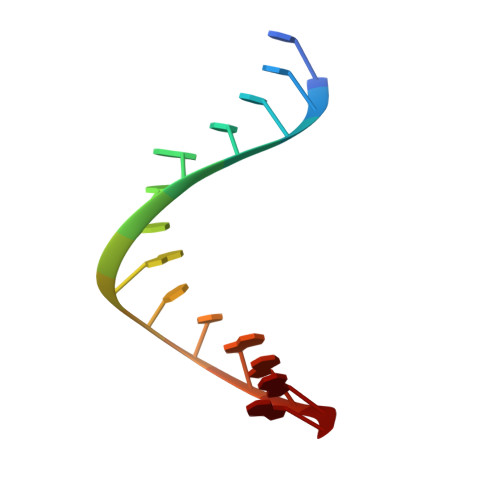
Structure of the tetradecanucleotide d(CCCCGGTACCGGGG)2 as an A-DNA duplex

Mandal, P.K.Venkadesh, S.Gautham, N.

(2012) Acta Crystallogr Sect F Struct Biol Cryst Commun 68: 393-399

  • DOI: https://doi.org/10.1107/S174430911200869X
  • Primary Citation Related Structures: 
    3V9D

  • PubMed Abstract: 

    The crystal structure of the tetradecanucleotide sequence d(CCCCGGTACCGGGG)(2) has been determined at 2.5 Å resolution in the tetragonal space group P4(1). This sequence was designed with the expectation of a four-way junction. However, the sequence crystallized as an A-DNA duplex and represents more than one full turn of the A-helix. The crystallographic asymmetric unit consists of one tetradecanucleotide duplex. The structural parameters of the A-type DNA duplex structure and the crystal-packing arrangement are described. One Mn(2+) ion was identified with direct coordination to the N7 position of G(13) and a water molecule at the major-groove side of the C(2)·G(13) base pair.


  • Organizational Affiliation
    • C. A. S. in Crystallography and Biophysics, University of Madras, Guindy, Chennai 600 025, India.

Macromolecule Content 

  • Total Structure Weight: 8.62 kDa 
  • Atom Count: 584 
  • Modeled Residue Count: 28 
  • Deposited Residue Count: 28 
  • Unique nucleic acid chains: 1

Macromolecules

Find similar nucleic acids by:  Sequence
Entity ID: 1
MoleculeChains LengthOrganismImage
DNA (5'-D(*CP*CP*CP*CP*GP*GP*TP*AP*CP*CP*GP*GP*GP*G)-3')
A, B
14N/A
Sequence Annotations
Expand
Reference Sequence

Small Molecules

Ligands 1 Unique
IDChains Name / Formula / InChI Key2D Diagram3D Interactions
MN

Query on MN


Download:Ideal Coordinates CCD File
C [auth B]MANGANESE (II) ION
Mn
WAEMQWOKJMHJLA-UHFFFAOYSA-N

Experimental Data & Validation

Experimental Data

  • Method: X-RAY DIFFRACTION
  • Resolution: 2.50 Å
  • R-Value Free:  0.244 (Depositor), 0.240 (DCC) 
  • R-Value Work:  0.217 (Depositor), 0.214 (DCC) 
  • R-Value Observed: 0.218 (Depositor) 
Space Group: P 41
Unit Cell:
Length ( Å )Angle ( ˚ )
a = 29.34α = 90
b = 29.34β = 90
c = 87.687γ = 90
Software Package:
Software NamePurpose
MAR345dtbdata collection
AMoREphasing
REFMACrefinement
AUTOMARdata reduction
SCALAdata scaling

Structure Validation

View Full Validation Report


Entry History 

Deposition Data

Revision History  (Full details and data files)

  • Version 1.0: 2012-04-18
    Type: Initial release
  • Version 1.1: 2014-03-12
    Changes: Database references
  • Version 1.2: 2024-03-20
    Changes: Data collection, Database references, Derived calculations
  • Version 1.3: 2024-04-03
    Changes: Refinement description