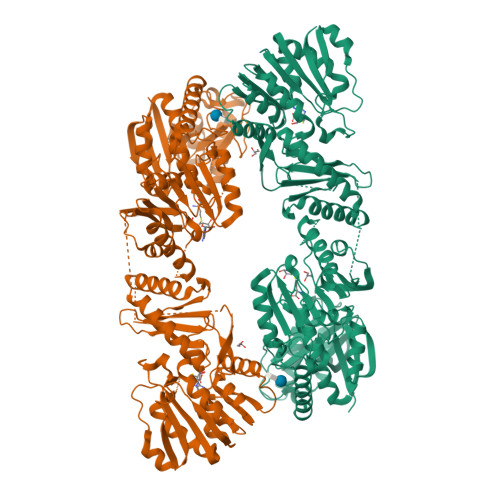
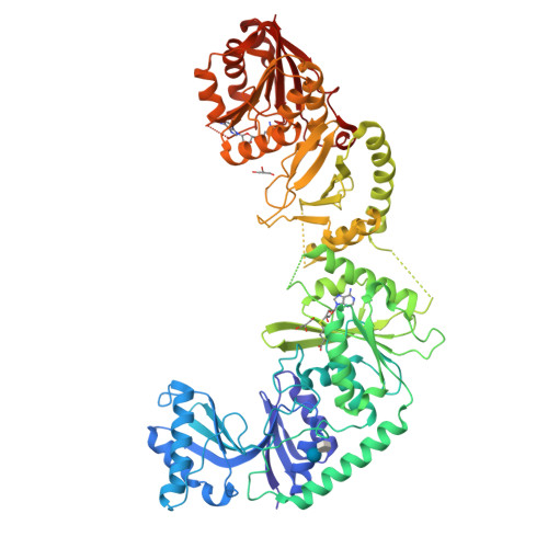
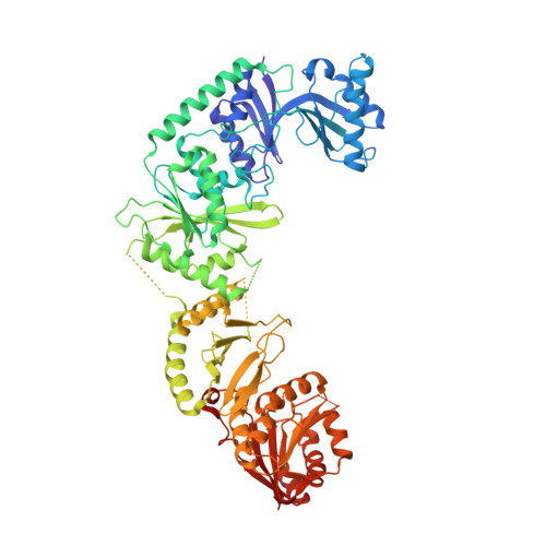
3V97

Crystal structure of bifunctional methyltransferase YcbY (RlmLK) from Escherichia coli, SAH binding


Experimental Data Snapshot

  • Method: X-RAY DIFFRACTION
  • Resolution: 2.20 Å
  • R-Value Free: 0.234 
  • R-Value Work: 0.186 
  • R-Value Observed: 0.189 

Starting Models: experimental
View more details

wwPDB Validation   3D Report Full Report


Ligand Structure Quality Assessment 


This is version 2.1 of the entry. See complete history


Literature

Structure of the bifunctional methyltransferase YcbY (RlmKL) that adds the m7G2069 and m2G2445 modifications in Escherichia coli 23S rRNA

Wang, K.T.Desmolaize, B.Nan, J.Zhang, X.W.Li, L.F.Douthwaite, S.Su, X.D.

(2012) Nucleic Acids Res 40: 5138-5148

  • DOI: https://doi.org/10.1093/nar/gks160
  • Primary Citation of Related Structures:  
    3V8V, 3V97

  • PubMed Abstract: 

    The 23S rRNA nucleotide m(2)G2445 is highly conserved in bacteria, and in Escherichia coli this modification is added by the enzyme YcbY. With lengths of around 700 amino acids, YcbY orthologs are the largest rRNA methyltransferases identified in Gram-negative bacteria, and they appear to be fusions from two separate proteins found in Gram-positives. The crystal structures described here show that both the N- and C-terminal halves of E. coli YcbY have a methyltransferase active site and their folding patterns respectively resemble the Streptococcus mutans proteins Smu472 and Smu776. Mass spectrometric analyses of 23S rRNAs showed that the N-terminal region of YcbY and Smu472 are functionally equivalent and add the m(2)G2445 modification, while the C-terminal region of YcbY is responsible for the m(7)G2069 methylation on the opposite side of the same helix (H74). Smu776 does not target G2069, and this nucleotide remains unmodified in Gram-positive rRNAs. The E.coli YcbY enzyme is the first example of a methyltransferase catalyzing two mechanistically different types of RNA modification, and has been renamed as the Ribosomal large subunit methyltransferase, RlmKL. Our structural and functional data provide insights into how this bifunctional enzyme evolved.


  • Organizational Affiliation

    State Key Laboratory of Protein and Plant Gene Research, and Biodynamic Optical Imaging Center (BIOPIC), School of Life Sciences, Peking University, Beijing 100871, Republic of China.


Macromolecules
Find similar proteins by:  (by identity cutoff)  |  3D Structure
Entity ID: 1
MoleculeChains Sequence LengthOrganismDetailsImage
Ribosomal RNA large subunit methyltransferase L
A, B
703Escherichia coli K-12Mutation(s): 0 
Gene Names: rlmL
EC: 2.1.1.173 (PDB Primary Data), 2.1.1.264 (UniProt)
UniProt
Find proteins for P75864 (Escherichia coli (strain K12))
Explore P75864 
Go to UniProtKB:  P75864
Entity Groups  
Sequence Clusters30% Identity50% Identity70% Identity90% Identity95% Identity100% Identity
UniProt GroupP75864
Sequence Annotations
Expand
  • Reference Sequence
Oligosaccharides

Help

Entity ID: 2
MoleculeChains Length2D Diagram Glycosylation3D Interactions
6-O-octanoyl-beta-D-fructofuranose-(2-1)-alpha-D-glucopyranose
C, D
2N/AN/A
Glycosylation Resources
GlyTouCan:  G98166HA
GlyCosmos:  G98166HA
Small Molecules
Ligands 2 Unique
IDChains Name / Formula / InChI Key2D Diagram3D Interactions
SAH
Query on SAH

Download Ideal Coordinates CCD File 
E [auth A],
F [auth A],
I [auth B],
J [auth B]
S-ADENOSYL-L-HOMOCYSTEINE
C14 H20 N6 O5 S
ZJUKTBDSGOFHSH-WFMPWKQPSA-N
GOL
Query on GOL

Download Ideal Coordinates CCD File 
G [auth A],
H [auth A],
K [auth B],
L [auth B]
GLYCEROL
C3 H8 O3
PEDCQBHIVMGVHV-UHFFFAOYSA-N
Biologically Interesting Molecules (External Reference) 1 Unique
Experimental Data & Validation

Experimental Data

  • Method: X-RAY DIFFRACTION
  • Resolution: 2.20 Å
  • R-Value Free: 0.234 
  • R-Value Work: 0.186 
  • R-Value Observed: 0.189 
  • Space Group: P 1 21 1
Unit Cell:
Length ( Å )Angle ( ˚ )
a = 73.646α = 90
b = 140.834β = 102.3
c = 102.866γ = 90
Software Package:
Software NamePurpose
MAR345dtbdata collection
MOLREPphasing
REFMACrefinement
HKL-2000data reduction
HKL-2000data scaling

Structure Validation

View Full Validation Report


Ligand Structure Quality Assessment 


Entry History 

Deposition Data

Revision History  (Full details and data files)

  • Version 1.0: 2012-02-29
    Type: Initial release
  • Version 1.1: 2013-06-26
    Changes: Database references
  • Version 2.0: 2020-07-29
    Type: Remediation
    Reason: Carbohydrate remediation
    Changes: Advisory, Atomic model, Data collection, Database references, Derived calculations, Non-polymer description, Structure summary
  • Version 2.1: 2023-11-08
    Changes: Data collection, Database references, Refinement description, Structure summary