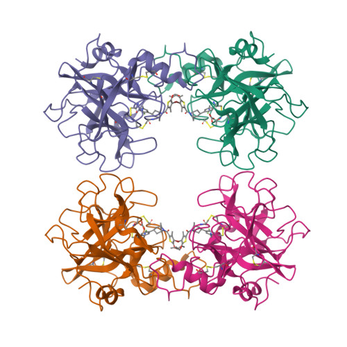
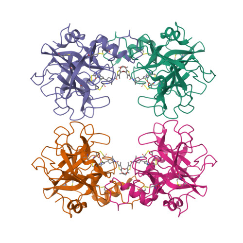
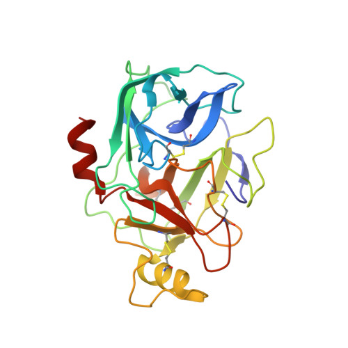
3V7T | pdb_00003v7t

Crystal Structure of Human Beta-Tryptase Complexed with a Synthetic Inhibitor with a Tropanylamide Scaffold


Experimental Data Snapshot

  • Method: X-RAY DIFFRACTION
  • Resolution: 2.09 Å
  • R-Value Free: 
    0.234 (Depositor), 0.240 (DCC) 
  • R-Value Work: 
    0.208 (Depositor), 0.210 (DCC) 
  • R-Value Observed: 
    0.210 (Depositor) 

wwPDB Validation   3D Report Full Report


Ligand Structure Quality Assessment 

Created with Raphaël 2.3.0Worse 01 BetterLigand structure goodness of fit to experimental dataBest fitted 0GXClick on this verticalbar to view details

This is version 1.2 of the entry. See complete history


Literature

A beta-tryptase inhibitor with a tropanylamide scaffold to improve in vitro stability and to lower hERG channel binding affinity

Liang, G.Choi-Sledeski, Y.M.Shum, P.Chen, X.Poli, G.B.Kumar, V.Minnich, A.Wang, Q.Tsay, J.Sides, K.Kang, J.Zhang, Y.

(2012) Bioorg Med Chem Lett 22: 1606-1610

  • DOI: https://doi.org/10.1016/j.bmcl.2011.12.127
  • Primary Citation of Related Structures:  
    3V7T

  • PubMed Abstract: 

    Tropanylamide was investigated as a possible scaffold for β-tryptase inhibitors with a basic benzylamine P1 group and a substituted thiophene P4 group. Comparing to piperidinylamide, the tropanylamide scaffold is much more rigid, which presents less opportunity for the inhibitor to bind with off-target proteins, such as cytochrome P450, SSAO, and hERG potassium channel. The proposed binding mode was further confirmed by an in-house X-ray structure through co-crystallization.


  • Organizational Affiliation

    Molecular Innovative Therapeutics, Sanofi Pharmaceuticals, Bridgewater, NJ 08807, USA. guyan.liang@sanofi-aventis.com


Macromolecules
Find similar proteins by:  (by identity cutoff)  |  3D Structure
Entity ID: 1
MoleculeChains Sequence LengthOrganismDetailsImage
TPSB2 protein
A, B, C, D
245Homo sapiensMutation(s): 0 
Gene Names: TPSB2
EC: 3.4.21.59
UniProt & NIH Common Fund Data Resources
Find proteins for P20231 (Homo sapiens)
Explore P20231 
Go to UniProtKB:  P20231
PHAROS:  P20231
GTEx:  ENSG00000197253 
Entity Groups  
Sequence Clusters30% Identity50% Identity70% Identity90% Identity95% Identity100% Identity
UniProt GroupP20231
Sequence Annotations
Expand
  • Reference Sequence
Experimental Data & Validation

Experimental Data

  • Method: X-RAY DIFFRACTION
  • Resolution: 2.09 Å
  • R-Value Free:  0.234 (Depositor), 0.240 (DCC) 
  • R-Value Work:  0.208 (Depositor), 0.210 (DCC) 
  • R-Value Observed: 0.210 (Depositor) 
Space Group: P 41
Unit Cell:
Length ( Å )Angle ( ˚ )
a = 81.719α = 90
b = 81.719β = 90
c = 170.684γ = 90
Software Package:
Software NamePurpose
DENZOdata reduction
SCALEPACKdata scaling
MOLREPphasing
BUSTER-TNTrefinement
PDB_EXTRACTdata extraction
HKL-2000data reduction
HKL-2000data scaling
BUSTERrefinement

Structure Validation

View Full Validation Report


Ligand Structure Quality Assessment 

Created with Raphaël 2.3.0Worse 01 BetterLigand structure goodness of fit to experimental dataBest fitted 0GXClick on this verticalbar to view details

Entry History 

Deposition Data

Revision History  (Full details and data files)

  • Version 1.0: 2012-03-14
    Type: Initial release
  • Version 1.1: 2017-11-08
    Changes: Refinement description
  • Version 1.2: 2024-11-27
    Changes: Data collection, Database references, Derived calculations, Structure summary