3V62 | pdb_00003v62

Structure of the S. cerevisiae Srs2 C-terminal domain in complex with PCNA conjugated to SUMO on lysine 164


Experimental Data Snapshot

  • Method: X-RAY DIFFRACTION
  • Resolution: 2.90 Å
  • R-Value Free: 
    0.237 (Depositor), 0.248 (DCC) 
  • R-Value Work: 
    0.184 (Depositor), 0.195 (DCC) 
  • R-Value Observed: 
    0.187 (Depositor) 

Starting Models: experimental
View more details

wwPDB Validation 3D Report Full Report

Validation slider image for 3V62

This is version 1.3 of the entry. See complete history

Literature

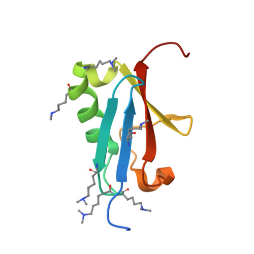
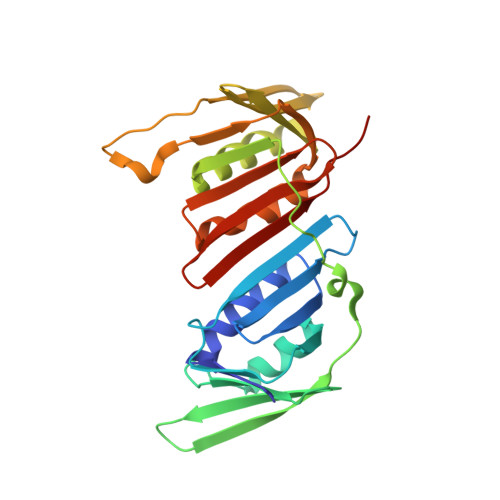
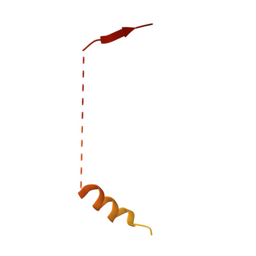
Recognition of SUMO-modified PCNA requires tandem receptor motifs in Srs2.

Armstrong, A.A.Mohideen, F.Lima, C.D.

(2012) Nature 483: 59-63

  • DOI: https://doi.org/10.1038/nature10883
  • Primary Citation Related Structures: 
    3V60, 3V61, 3V62

  • PubMed Abstract: 

    Ubiquitin (Ub) and ubiquitin-like (Ubl) modifiers such as SUMO (also known as Smt3 in Saccharomyces cerevisiae) mediate signal transduction through post-translational modification of substrate proteins in pathways that control differentiation, apoptosis and the cell cycle, and responses to stress such as the DNA damage response. In yeast, the proliferating cell nuclear antigen PCNA (also known as Pol30) is modified by ubiquitin in response to DNA damage and by SUMO during S phase. Whereas Ub-PCNA can signal for recruitment of translesion DNA polymerases, SUMO-PCNA signals for recruitment of the anti-recombinogenic DNA helicase Srs2. It remains unclear how receptors such as Srs2 specifically recognize substrates after conjugation to Ub and Ubls. Here we show, through structural, biochemical and functional studies, that the Srs2 carboxy-terminal domain harbours tandem receptor motifs that interact independently with PCNA and SUMO and that both motifs are required to recognize SUMO-PCNA specifically. The mechanism presented is pertinent to understanding how other receptors specifically recognize Ub- and Ubl-modified substrates to facilitate signal transduction.


  • Organizational Affiliation
    • Structural Biology Program, Sloan-Kettering Institute, New York, New York 10065, USA.

Macromolecule Content 

  • Total Structure Weight: 93.8 kDa 
  • Atom Count: 5,723 
  • Modeled Residue Count: 710 
  • Deposited Residue Count: 822 
  • Unique protein chains: 3

Macromolecules

Find similar proteins by:|  3D Structure
Entity ID: 1
MoleculeChains  Sequence LengthOrganismDetailsImage
Ubiquitin-like protein SMT3
A, D
84Saccharomyces cerevisiae S288CMutation(s): 0 
Gene Names: D9719.15SMT3YDR510W
UniProt
Find proteins for Q12306 (Saccharomyces cerevisiae (strain ATCC 204508 / S288c))
Explore Q12306 
Go to UniProtKB:  Q12306
Entity Groups
Sequence Clusters30% Identity50% Identity70% Identity90% Identity95% Identity100% Identity
UniProt GroupQ12306
Sequence Annotations
Expand
Reference Sequence
Find similar proteins by:|  3D Structure
Entity ID: 2
MoleculeChains  Sequence LengthOrganismDetailsImage
Proliferating cell nuclear antigen
B, E
258Saccharomyces cerevisiae S288CMutation(s): 1 
Gene Names: POL30YBR0811YBR088C
UniProt
Find proteins for P15873 (Saccharomyces cerevisiae (strain ATCC 204508 / S288c))
Explore P15873 
Go to UniProtKB:  P15873
Entity Groups
Sequence Clusters30% Identity50% Identity70% Identity90% Identity95% Identity100% Identity
UniProt GroupP15873
Sequence Annotations
Expand
Reference Sequence
Find similar proteins by:|  3D Structure
Entity ID: 3
MoleculeChains  Sequence LengthOrganismDetailsImage
ATP-dependent DNA helicase SRS2
C, F
69Saccharomyces cerevisiae S288CMutation(s): 0 
Gene Names: HPR5J0913RADHSRS2YJL092W
EC: 3.6.4.12 (PDB Primary Data), 5.6.2.4 (UniProt)
UniProt
Find proteins for P12954 (Saccharomyces cerevisiae (strain ATCC 204508 / S288c))
Explore P12954 
Go to UniProtKB:  P12954
Entity Groups
Sequence Clusters30% Identity50% Identity70% Identity90% Identity95% Identity100% Identity
UniProt GroupP12954
Sequence Annotations
Expand
Reference Sequence

Small Molecules

Ligands 2 Unique
IDChains Name / Formula / InChI Key2D Diagram3D Interactions
NEQ

Query on NEQ


Download:Ideal Coordinates CCD File
G [auth B],
H [auth B],
K [auth E],
L [auth E]
N-ETHYLMALEIMIDE
C6 H7 N O2
HDFGOPSGAURCEO-UHFFFAOYSA-N
SO4

Query on SO4


Download:Ideal Coordinates CCD File
I [auth B],
J [auth B],
M [auth E],
N [auth E]
SULFATE ION
O4 S
QAOWNCQODCNURD-UHFFFAOYSA-L
Modified Residues  1 Unique
IDChains TypeFormula2D DiagramParent
MLY
Query on MLY
A, D
L-PEPTIDE LINKINGC8 H18 N2 O2LYS

Experimental Data & Validation

Experimental Data

  • Method: X-RAY DIFFRACTION
  • Resolution: 2.90 Å
  • R-Value Free:  0.237 (Depositor), 0.248 (DCC) 
  • R-Value Work:  0.184 (Depositor), 0.195 (DCC) 
  • R-Value Observed: 0.187 (Depositor) 
Space Group: C 1 2 1
Unit Cell:
Length ( Å )Angle ( ˚ )
a = 196.817α = 90
b = 62.256β = 135.04
c = 139.247γ = 90
Software Package:
Software NamePurpose
DENZOdata reduction
SCALEPACKdata scaling
REFMACrefinement
PDB_EXTRACTdata extraction
ADSCdata collection
HKL-2000data reduction
PHASERphasing

Structure Validation

View Full Validation Report


Entry History 

Deposition Data

Revision History  (Full details and data files)

  • Version 1.0: 2012-02-29
    Type: Initial release
  • Version 1.1: 2013-04-03
    Changes: Database references
  • Version 1.2: 2017-10-25
    Changes: Author supporting evidence, Refinement description
  • Version 1.3: 2023-09-13
    Changes: Data collection, Database references, Derived calculations, Refinement description