


Crystal structure of the mutant E234A of Galacturonate Dehydratase from GEOBACILLUS SP. complexed with Mg
Fedorov, A.A., Fedorov, E.V., Groninger-Poe, F., Gerlt, J.A., Almo, S.C.To be published.
Experimental Data Snapshot
Starting Model: experimental
View more details
wwPDB Validation 3D Report Full Report

Entity ID: 1 | |||||
|---|---|---|---|---|---|
| Molecule | Chains | Sequence Length | Organism | Details | Image |
| Mandelate racemase/muconate lactonizing protein | 392 | Paenibacillus sp. Y412MC10 | Mutation(s): 1 Gene Names: GYMC10_3367 | 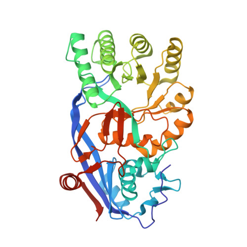 | |
| Ligands 2 Unique | |||||
|---|---|---|---|---|---|
| ID | Chains | Name / Formula / InChI Key | 2D Diagram | 3D Interactions | |
| EPE Download:Ideal Coordinates CCD File | D [auth A] | 4-(2-HYDROXYETHYL)-1-PIPERAZINE ETHANESULFONIC ACID C8 H18 N2 O4 S JKMHFZQWWAIEOD-UHFFFAOYSA-N |  | ||
| MG Download:Ideal Coordinates CCD File | B [auth A], C [auth A] | MAGNESIUM ION Mg JLVVSXFLKOJNIY-UHFFFAOYSA-N |  | ||
| Length ( Å ) | Angle ( ˚ ) |
|---|---|
| a = 105.986 | α = 90 |
| b = 122.853 | β = 90 |
| c = 134.263 | γ = 90 |
| Software Name | Purpose |
|---|---|
| CBASS | data collection |
| BALBES | phasing |
| PHENIX | refinement |
| HKL-2000 | data reduction |
| SCALEPACK | data scaling |


