3V4Y | pdb_00003v4y

Crystal Structure of the first Nuclear PP1 holoenzyme


Experimental Data Snapshot

  • Method: X-RAY DIFFRACTION
  • Resolution: 2.10 Å
  • R-Value Free: 
    0.196 (Depositor), 0.190 (DCC) 
  • R-Value Work: 
    0.154 (Depositor), 0.152 (DCC) 
  • R-Value Observed: 
    0.156 (Depositor) 

wwPDB Validation 3D Report Full Report

Validation slider image for 3V4Y

This is version 1.1 of the entry. See complete history

Literature

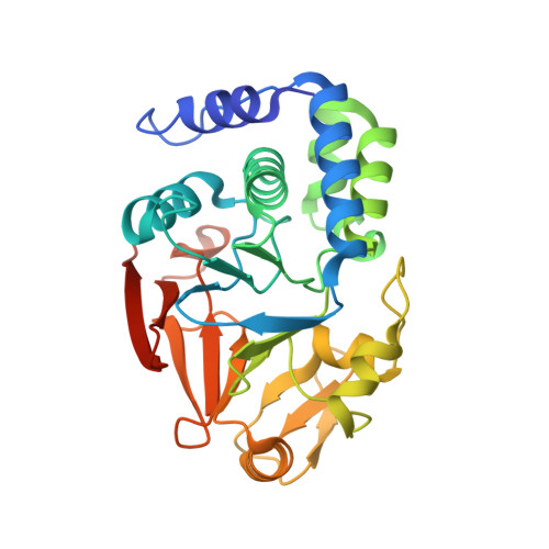
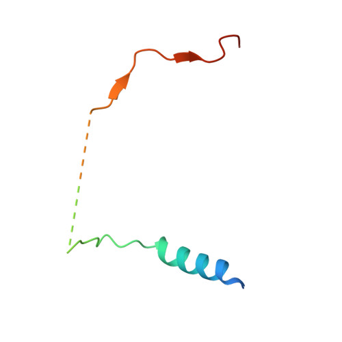
The Molecular Basis for Substrate Specificity of the Nuclear NIPP1:PP1 Holoenzyme.

O'Connell, N.Nichols, S.R.Heroes, E.Beullens, M.Bollen, M.Peti, W.Page, R.

(2012) Structure 20: 1746-1756

  • DOI: https://doi.org/10.1016/j.str.2012.08.003
  • Primary Citation Related Structures: 
    3V4Y

  • PubMed Abstract: 

    Regulation of protein phosphatase 1 (PP1) is controlled by a diverse array of regulatory proteins. However, how these proteins direct PP1 specificity is not well understood. More than one-third of the nuclear pool of PP1 forms a holoenzyme with the nuclear inhibitor of PP1, NIPP1, to regulate chromatin remodeling, among other essential biological functions. Here, we show that the PP1-binding domain of NIPP1 is an intrinsically disordered protein, which binds PP1 in an unexpected manner. NIPP1 forms an α helix that engages PP1 at a unique interaction site, using polar rather than hydrophobic contacts. Importantly, the structure also reveals a shared PP1 interaction site outside of the RVxF motif, the ΦΦ motif. Finally, we show that NIPP1:PP1 substrate selectivity is determined by altered electrostatics and enhanced substrate localization. Together, our results provide the molecular basis by which NIPP1 directs PP1 substrate specificity in the nucleus.


  • Organizational Affiliation
    • Department of Molecular Pharmacology, Physiology and Biotechnology, Brown University, Providence, RI 02912, USA.

Macromolecule Content 

  • Total Structure Weight: 171.51 kDa 
  • Atom Count: 11,550 
  • Modeled Residue Count: 1,340 
  • Deposited Residue Count: 1,472 
  • Unique protein chains: 2

Macromolecules

Find similar proteins by:|  3D Structure
Entity ID: 1
MoleculeChains  Sequence LengthOrganismDetailsImage
Serine/threonine-protein phosphatase PP1-alpha catalytic subunit
A, C, E, G
306Homo sapiensMutation(s): 0 
Gene Names: PPP1APPP1CA
EC: 3.1.3.16
UniProt & NIH Common Fund Data Resources
Find proteins for P62136 (Homo sapiens)
Explore P62136 
Go to UniProtKB:  P62136
PHAROS:  P62136
GTEx:  ENSG00000172531 
Entity Groups
Sequence Clusters30% Identity50% Identity70% Identity90% Identity95% Identity100% Identity
UniProt GroupP62136
Sequence Annotations
Expand
Reference Sequence
Find similar proteins by:|  3D Structure
Entity ID: 2
MoleculeChains  Sequence LengthOrganismDetailsImage
Nuclear inhibitor of protein phosphatase 1
B, D, F, H
62Homo sapiensMutation(s): 0 
Gene Names: PPP1R8ARD1NIPP1
EC: 3.1.4
UniProt & NIH Common Fund Data Resources
Find proteins for Q12972 (Homo sapiens)
Explore Q12972 
Go to UniProtKB:  Q12972
PHAROS:  Q12972
GTEx:  ENSG00000117751 
Entity Groups
Sequence Clusters30% Identity50% Identity70% Identity90% Identity95% Identity100% Identity
UniProt GroupQ12972
Sequence Annotations
Expand
Reference Sequence

Small Molecules

Ligands 3 Unique
IDChains Name / Formula / InChI Key2D Diagram3D Interactions
15P

Query on 15P


Download:Ideal Coordinates CCD File
N [auth A]POLYETHYLENE GLYCOL (N=34)
C69 H140 O35
VUYXVWGKCKTUMF-UHFFFAOYSA-N
GOL

Query on GOL


Download:Ideal Coordinates CCD File
AA [auth G]
K [auth A]
L [auth A]
M [auth A]
Q [auth C]
AA [auth G],
K [auth A],
L [auth A],
M [auth A],
Q [auth C],
R [auth C],
S [auth C],
V [auth E],
W [auth E],
X [auth E]
GLYCEROL
C3 H8 O3
PEDCQBHIVMGVHV-UHFFFAOYSA-N
MN

Query on MN


Download:Ideal Coordinates CCD File
I [auth A]
J [auth A]
O [auth C]
P [auth C]
T [auth E]
I [auth A],
J [auth A],
O [auth C],
P [auth C],
T [auth E],
U [auth E],
Y [auth G],
Z [auth G]
MANGANESE (II) ION
Mn
WAEMQWOKJMHJLA-UHFFFAOYSA-N

Experimental Data & Validation

Experimental Data

  • Method: X-RAY DIFFRACTION
  • Resolution: 2.10 Å
  • R-Value Free:  0.196 (Depositor), 0.190 (DCC) 
  • R-Value Work:  0.154 (Depositor), 0.152 (DCC) 
  • R-Value Observed: 0.156 (Depositor) 
Space Group: P 21 21 21
Unit Cell:
Length ( Å )Angle ( ˚ )
a = 76.639α = 90
b = 116.035β = 90
c = 168.093γ = 90
Software Package:
Software NamePurpose
CBASSdata collection
PHENIXmodel building
PHENIXrefinement
HKL-2000data reduction
HKL-2000data scaling
PHENIXphasing

Structure Validation

View Full Validation Report


Entry History 

Deposition Data

Revision History  (Full details and data files)

  • Version 1.0: 2012-10-31
    Type: Initial release
  • Version 1.1: 2024-02-28
    Changes: Data collection, Database references, Derived calculations