3V3C | pdb_00003v3c

Crystal Structure of Chloroplast ATP synthase c-ring from Pisum sativum


Experimental Data Snapshot

  • Method: X-RAY DIFFRACTION
  • Resolution: 3.40 Å
  • R-Value Free: 
    0.322 (Depositor), 0.330 (DCC) 
  • R-Value Work: 
    0.299 (Depositor), 0.300 (DCC) 
  • R-Value Observed: 
    0.301 (Depositor) 

wwPDB Validation   3D Report Full Report


Ligand Structure Quality Assessment 

Created with Raphaël 2.3.0Worse 01 BetterLigand structure goodness of fit to experimental dataBest fitted DGDClick on this verticalbar to view details

This is version 1.2 of the entry. See complete history


Literature

Structure and flexibility of the C-ring in the electromotor of rotary F(o)F(1)-ATPase of pea chloroplasts.

Saroussi, S.Schushan, M.Ben-Tal, N.Junge, W.Nelson, N.

(2012) PLoS One 7: e43045-e43045

  • DOI: https://doi.org/10.1371/journal.pone.0043045
  • Primary Citation of Related Structures:  
    3V3C

  • PubMed Abstract: 

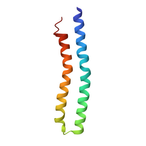
    A ring of 8-15 identical c-subunits is essential for ion-translocation by the rotary electromotor of the ubiquitous F(O)F(1)-ATPase. Here we present the crystal structure at 3.4Å resolution of the c-ring from chloroplasts of a higher plant (Pisum sativum), determined using a native preparation. The crystal structure was found to resemble that of an (ancestral) cyanobacterium. Using elastic network modeling to investigate the ring's eigen-modes, we found five dominant modes of motion that fell into three classes. They revealed the following deformations of the ring: (I) ellipsoidal, (II) opposite twisting of the luminal circular surface of the ring against the stromal surface, and (III) kinking of the hairpin-shaped monomers in the middle, resulting in bending/stretching of the ring. Extension of the elastic network analysis to rings of different c(n)-symmetry revealed the same classes of dominant modes as in P. sativum (c(14)). We suggest the following functional roles for these classes: The first and third classes of modes affect the interaction of the c-ring with its counterparts in F(O), namely subunits a and bb'. These modes are likely to be involved in ion-translocation and torque generation. The second class of deformation, along with deformations of subunits γ and ε might serve to elastically buffer the torque transmission between F(O) and F(1).


  • Organizational Affiliation

    Department of Biochemistry and Molecular Biology, George S. Wise Faculty of Life Sciences, Tel-Aviv University, Ramat Aviv, Israel.


Macromolecules
Find similar proteins by:  (by identity cutoff)  |  3D Structure
Entity ID: 1
MoleculeChains Sequence LengthOrganismDetailsImage
ATP synthase subunit c, chloroplastic
A, B, C, D, E
79Pisum sativumMutation(s): 0 
Membrane Entity: Yes 
UniProt
Find proteins for P08212 (Pisum sativum)
Explore P08212 
Go to UniProtKB:  P08212
Entity Groups  
Sequence Clusters30% Identity50% Identity70% Identity90% Identity95% Identity100% Identity
UniProt GroupP08212
Sequence Annotations
Expand
  • Reference Sequence
Small Molecules
Ligands 3 Unique
IDChains Name / Formula / InChI Key2D Diagram3D Interactions
DGD
Query on DGD

Download Ideal Coordinates CCD File 
O [auth A]DIGALACTOSYL DIACYL GLYCEROL (DGDG)
C51 H96 O15
LDQFLSUQYHBXSX-HXXRYREZSA-N
YT3
Query on YT3

Download Ideal Coordinates CCD File 
BA [auth G]
DA [auth H]
HA [auth K]
JA [auth L]
MA [auth N]
YTTRIUM (III) ION
Y
GRTBAGCGDOYUBE-UHFFFAOYSA-N
NA
Query on NA

Download Ideal Coordinates CCD File 
AA [auth F]
CA [auth G]
EA [auth H]
FA [auth I]
GA [auth J]
SODIUM ION
Na
FKNQFGJONOIPTF-UHFFFAOYSA-N
Experimental Data & Validation

Experimental Data

  • Method: X-RAY DIFFRACTION
  • Resolution: 3.40 Å
  • R-Value Free:  0.322 (Depositor), 0.330 (DCC) 
  • R-Value Work:  0.299 (Depositor), 0.300 (DCC) 
  • R-Value Observed: 0.301 (Depositor) 
Space Group: C 1 2 1
Unit Cell:
Length ( Å )Angle ( ˚ )
a = 139.13α = 90
b = 102.38β = 101.22
c = 122.43γ = 90
Software Package:
Software NamePurpose
SCALAdata scaling
PHASERphasing
PHENIXrefinement
PDB_EXTRACTdata extraction
XDSdata scaling
XDSdata reduction

Structure Validation

View Full Validation Report


Ligand Structure Quality Assessment 

Created with Raphaël 2.3.0Worse 01 BetterLigand structure goodness of fit to experimental dataBest fitted DGDClick on this verticalbar to view details

Entry History 

Deposition Data

Revision History  (Full details and data files)

  • Version 1.0: 2012-10-03
    Type: Initial release
  • Version 1.1: 2013-01-23
    Changes: Database references
  • Version 1.2: 2024-02-28
    Changes: Data collection, Database references, Derived calculations, Refinement description