3UXU | pdb_00003uxu

The structure of the catalytic domain of the Sulfolobus Spindle-shaped viral integrase reveals an evolutionarily conserved catalytic core and supports a mechanism of DNA cleavage in trans


Experimental Data Snapshot

  • Method: X-RAY DIFFRACTION
  • Resolution: 2.71 Å
  • R-Value Free: 
    0.214 (Depositor), 0.227 (DCC) 
  • R-Value Work: 
    0.193 (Depositor), 0.206 (DCC) 
  • R-Value Observed: 
    0.194 (Depositor) 

wwPDB Validation 3D Report Full Report

Validation slider image for 3UXU

This is version 1.3 of the entry. See complete history

Literature

The Structure of an Archaeal Viral Integrase Reveals an Evolutionarily Conserved Catalytic Core yet Supports a Mechanism of DNA Cleavage in trans.

Eilers, B.J.Young, M.J.Lawrence, C.M.

(2012) J Virol 86: 8309-8313

  • DOI: https://doi.org/10.1128/JVI.00547-12
  • Primary Citation Related Structures: 
    3UXU

  • PubMed Abstract: 

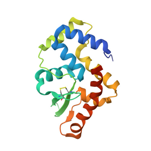
    The first structure of a catalytic domain from a hyperthermophilic archaeal viral integrase reveals a minimal fold similar to that of bacterial HP1 integrase and defines structural elements conserved across three domains of life. However, structural superposition on bacterial Holliday junction complexes and similarities in the C-terminal tail with that of eukaryotic Flp suggest that the catalytic tyrosine and an additional active-site lysine are delivered to neighboring subunits in trans. An intramolecular disulfide bond contributes significant thermostability in vitro.


  • Organizational Affiliation
    • Thermal Biology Institute, Montana State University, Bozeman, Montana, USA.

Macromolecule Content 

  • Total Structure Weight: 20.12 kDa 
  • Atom Count: 1,367 
  • Modeled Residue Count: 159 
  • Deposited Residue Count: 169 
  • Unique protein chains: 1

Macromolecules

Find similar proteins by:|  3D Structure
Entity ID: 1
MoleculeChains  Sequence LengthOrganismDetailsImage
Probable integrase169Sulfolobus spindle-shaped virus 1Mutation(s): 2 
Gene Names: d335
EC: 3.1 (UniProt), 2.7.7 (UniProt)
UniProt
Find proteins for P20214 (Sulfolobus spindle-shape virus 1)
Explore P20214 
Go to UniProtKB:  P20214
Entity Groups
Sequence Clusters30% Identity50% Identity70% Identity90% Identity95% Identity100% Identity
UniProt GroupP20214
Sequence Annotations
Expand
Reference Sequence

Small Molecules

Ligands 1 Unique
IDChains Name / Formula / InChI Key2D Diagram3D Interactions
PO4

Query on PO4


Download:Ideal Coordinates CCD File
B [auth A]PHOSPHATE ION
O4 P
NBIIXXVUZAFLBC-UHFFFAOYSA-K

Experimental Data & Validation

Experimental Data

  • Method: X-RAY DIFFRACTION
  • Resolution: 2.71 Å
  • R-Value Free:  0.214 (Depositor), 0.227 (DCC) 
  • R-Value Work:  0.193 (Depositor), 0.206 (DCC) 
  • R-Value Observed: 0.194 (Depositor) 
Space Group: P 65 2 2
Unit Cell:
Length ( Å )Angle ( ˚ )
a = 73.963α = 90
b = 73.963β = 90
c = 176.251γ = 120
Software Package:
Software NamePurpose
Blu-Icedata collection
AutoSolphasing
REFMACrefinement
HKL-2000data reduction
SCALEPACKdata scaling

Structure Validation

View Full Validation Report


Entry History 

Deposition Data

Revision History  (Full details and data files)

  • Version 1.0: 2012-05-16
    Type: Initial release
  • Version 1.1: 2012-07-25
    Changes: Database references
  • Version 1.2: 2017-11-08
    Changes: Refinement description
  • Version 1.3: 2024-10-16
    Changes: Data collection, Database references, Derived calculations, Structure summary