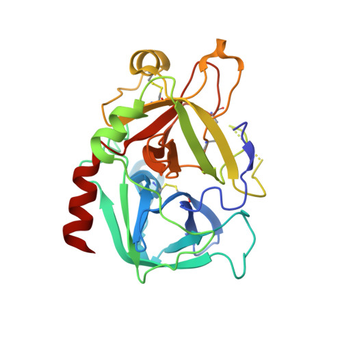
3UTU

High affinity inhibitor of human thrombin


Experimental Data Snapshot

  • Method: X-RAY DIFFRACTION
  • Resolution: 1.55 Å
  • R-Value Free: 0.271 
  • R-Value Work: 0.217 
  • R-Value Observed: 0.244 

Starting Model: experimental
View more details

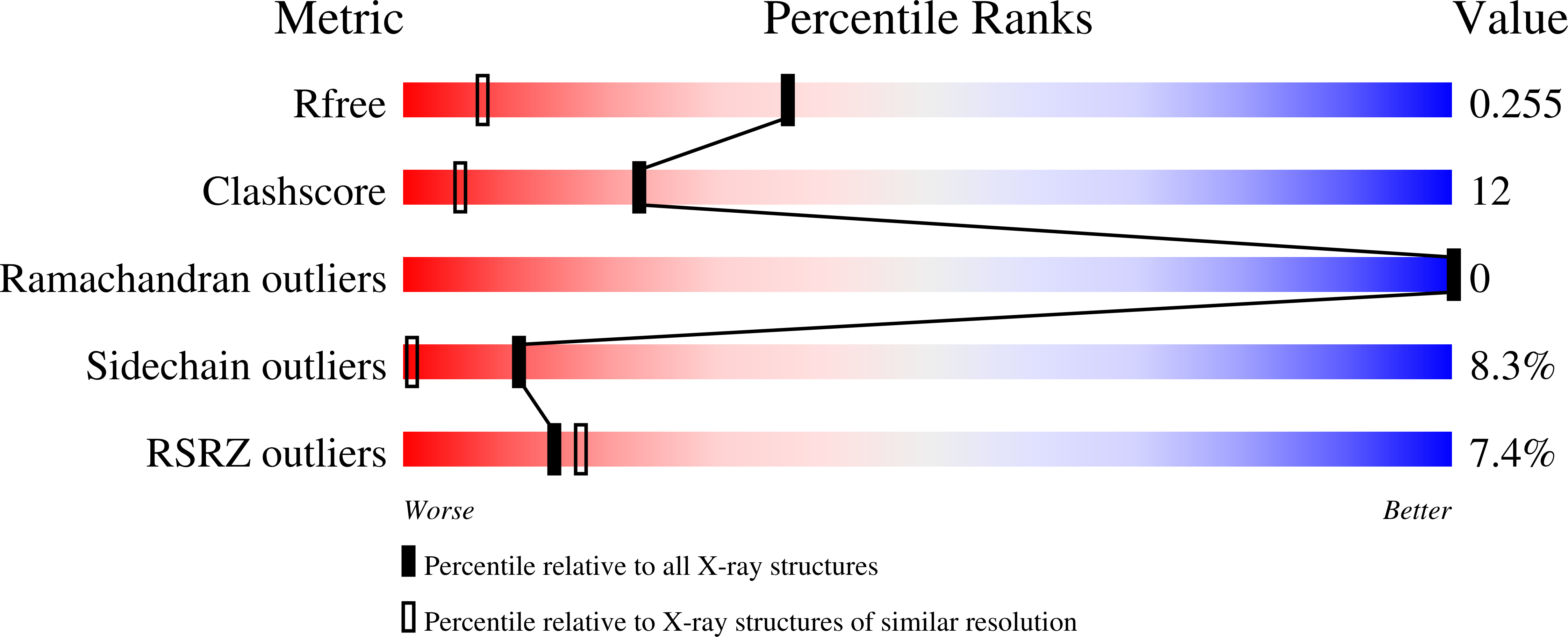
wwPDB Validation   3D Report Full Report


Ligand Structure Quality Assessment 


This is version 1.6 of the entry. See complete history


Literature

Beyond heparinization: design of highly potent thrombin inhibitors suitable for surface coupling

Steinmetzer, T.Baum, B.Biela, A.Klebe, G.Nowak, G.Bucha, E.

(2012) ChemMedChem 7: 1965-1973

  • DOI: https://doi.org/10.1002/cmdc.201200292
  • Primary Citation of Related Structures:  
    3UTU

  • PubMed Abstract: 

    During extracorporeal circulation, when blood comes in contact with artificial surfaces, patients receive a standard treatment with anticoagulants to avoid blood coagulation. Dialysis patients in particular are systemically treated with heparin up to four times a week, causing a high burden for the body. For potential anticoagulant modification of external materials, such as dialysis equipment, a series of highly potent thrombin inhibitors was developed. All inhibitors share the general formula arylsulfonyl-P3-Pro-4-amidinobenzylamide, where P3 is glycyl or a trifunctional amino acid residue in L-configuration. Among this series, several derivatives inhibit thrombin with Ki values of less than 1 nM. Specificity measurements revealed that this inhibitor type is highly specific for thrombin with negligible activity against related trypsin-like serine proteases. X-ray analysis of the most potent analogue in complex with thrombin demonstrated that the N-terminal arylsulfonyl group occupies the aryl binding site, whereas the P3 side chain is directed into the solvent and therefore is well suited for further coupling. Based on their in vitro profile, these inhibitors are suitable candidates for the development of hemocompatible materials with anticoagulant properties.


  • Organizational Affiliation

    Institute of Pharmaceutical Chemistry, Philipps University, Marbacher Weg 6, 35032 Marburg (Germany). steinmetzer@staff.uni-marburg.de.


Macromolecules
Find similar proteins by:  (by identity cutoff)  |  3D Structure
Entity ID: 1
MoleculeChains Sequence LengthOrganismDetailsImage
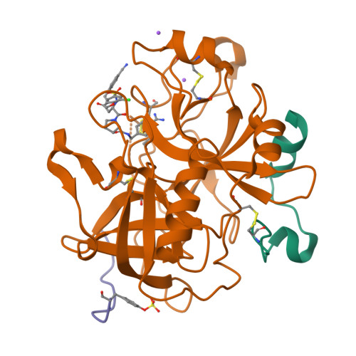
Thrombin light chainA [auth L]36Homo sapiensMutation(s): 0 
EC: 3.4.21.5
UniProt & NIH Common Fund Data Resources
Find proteins for P00734 (Homo sapiens)
Explore P00734 
Go to UniProtKB:  P00734
PHAROS:  P00734
GTEx:  ENSG00000180210 
Entity Groups  
Sequence Clusters30% Identity50% Identity70% Identity90% Identity95% Identity100% Identity
UniProt GroupP00734
Sequence Annotations
Expand
  • Reference Sequence
Find similar proteins by:  (by identity cutoff)  |  3D Structure
Entity ID: 2
MoleculeChains Sequence LengthOrganismDetailsImage
Thrombin heavy chainB [auth H]259Homo sapiensMutation(s): 0 
EC: 3.4.21.5
UniProt & NIH Common Fund Data Resources
Find proteins for P00734 (Homo sapiens)
Explore P00734 
Go to UniProtKB:  P00734
PHAROS:  P00734
GTEx:  ENSG00000180210 
Entity Groups  
Sequence Clusters30% Identity50% Identity70% Identity90% Identity95% Identity100% Identity
UniProt GroupP00734
Sequence Annotations
Expand
  • Reference Sequence

Find similar proteins by:  Sequence   |   3D Structure  

Entity ID: 3
MoleculeChains Sequence LengthOrganismDetailsImage
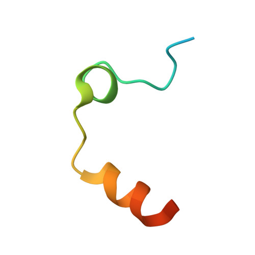
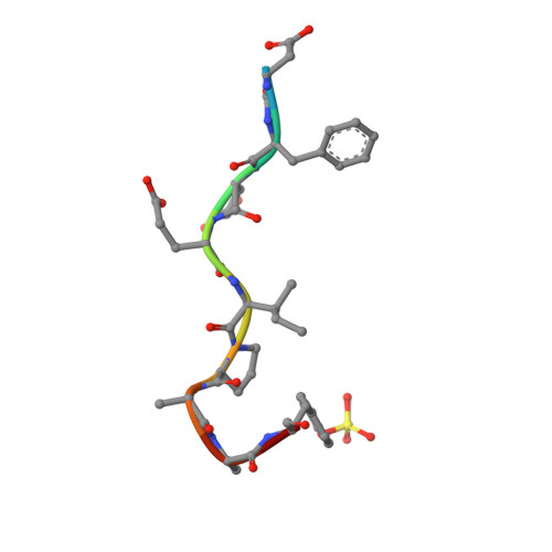
Hirudin variant-1C [auth I]11Hirudo medicinalisMutation(s): 0 
UniProt
Find proteins for P01050 (Hirudo medicinalis)
Explore P01050 
Go to UniProtKB:  P01050
Entity Groups  
Sequence Clusters30% Identity50% Identity70% Identity90% Identity95% Identity100% Identity
UniProt GroupP01050
Sequence Annotations
Expand
  • Reference Sequence
Small Molecules
Ligands 2 Unique
IDChains Name / Formula / InChI Key2D Diagram3D Interactions
1TS
Query on 1TS

Download Ideal Coordinates CCD File 
D [auth H](2S)-N-[(4-carbamimidoylphenyl)methyl]-1-[(2S)-2-[(3-chloro-4-methoxybenzene)sulfonamido]-3-{[(4-cyanophenyl)methyl]carbamoyl}propanoyl]pyrrolidine-2-carboxamide
C32 H34 Cl N7 O6 S
YNEVNNWCKYNFGX-SVBPBHIXSA-N
NA
Query on NA

Download Ideal Coordinates CCD File 
E [auth H],
F [auth H]
SODIUM ION
Na
FKNQFGJONOIPTF-UHFFFAOYSA-N
Modified Residues  1 Unique
IDChains TypeFormula2D DiagramParent
TYS
Query on TYS
C [auth I]L-PEPTIDE LINKINGC9 H11 N O6 STYR
Binding Affinity Annotations 
IDSourceBinding Affinity
1TS PDBBind:  3UTU Ki: 0.01 (nM) from 1 assay(s)
Experimental Data & Validation

Experimental Data

  • Method: X-RAY DIFFRACTION
  • Resolution: 1.55 Å
  • R-Value Free: 0.271 
  • R-Value Work: 0.217 
  • R-Value Observed: 0.244 
  • Space Group: C 1 2 1
Unit Cell:
Length ( Å )Angle ( ˚ )
a = 70.77α = 90
b = 71.35β = 100.65
c = 72.39γ = 90
Software Package:
Software NamePurpose
HKL-2000data collection
SHELXmodel building
SHELXL-97refinement
HKL-2000data reduction
HKL-2000data scaling
SHELXphasing

Structure Validation

View Full Validation Report


Ligand Structure Quality Assessment 


Entry History 

Deposition Data

Revision History  (Full details and data files)

  • Version 1.0: 2012-08-29
    Type: Initial release
  • Version 1.1: 2012-12-12
    Changes: Other
  • Version 1.2: 2013-03-13
    Changes: Other
  • Version 1.3: 2014-03-12
    Changes: Database references
  • Version 1.4: 2023-11-08
    Changes: Data collection, Database references, Derived calculations, Refinement description
  • Version 1.5: 2023-12-06
    Changes: Data collection
  • Version 1.6: 2024-11-20
    Changes: Structure summary