3USE | pdb_00003use

Crystal Structure of E. coli hydrogenase-1 in its as-isolated form


Experimental Data Snapshot

  • Method: X-RAY DIFFRACTION
  • Resolution: 1.67 Å
  • R-Value Free: 
    0.168 (Depositor), 0.170 (DCC) 
  • R-Value Work: 
    0.120 (Depositor), 0.121 (DCC) 
  • R-Value Observed: 
    0.122 (Depositor) 

Starting Model: experimental
View more details

wwPDB Validation 3D Report Full Report

Validation slider image for 3USE

Ligand Structure Quality Assessment 


This is version 1.2 of the entry. See complete history

Literature

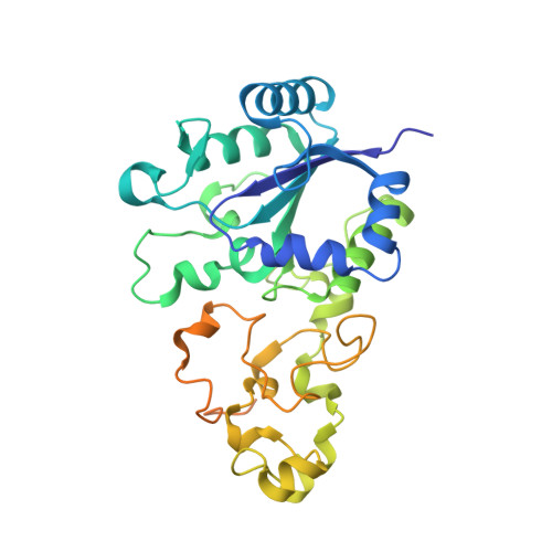
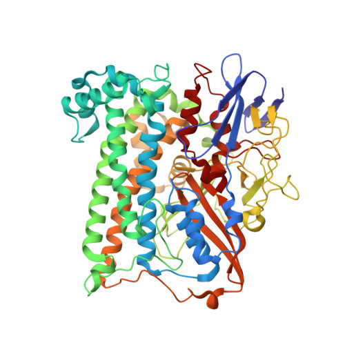
X-ray crystallographic and computational studies of the O2-tolerant [NiFe]-hydrogenase 1 from Escherichia coli.

Volbeda, A.Amara, P.Darnault, C.Mouesca, J.M.Parkin, A.Roessler, M.M.Armstrong, F.A.Fontecilla-Camps, J.C.

(2012) Proc Natl Acad Sci U S A 109: 5305-5310

  • DOI: https://doi.org/10.1073/pnas.1119806109
  • Primary Citation Related Structures: 
    3UQY, 3USC, 3USE

  • PubMed Abstract: 

    The crystal structure of the membrane-bound O(2)-tolerant [NiFe]-hydrogenase 1 from Escherichia coli (EcHyd-1) has been solved in three different states: as-isolated, H(2)-reduced, and chemically oxidized. As very recently reported for similar enzymes from Ralstonia eutropha and Hydrogenovibrio marinus, two supernumerary Cys residues coordinate the proximal [FeS] cluster in EcHyd-1, which lacks one of the inorganic sulfide ligands. We find that the as-isolated, aerobically purified species contains a mixture of at least two conformations for one of the cluster iron ions and Glu76. In one of them, Glu76 and the iron occupy positions that are similar to those found in O(2)-sensitive [NiFe]-hydrogenases. In the other conformation, this iron binds, besides three sulfur ligands, the amide N from Cys20 and one Oε of Glu76. Our calculations show that oxidation of this unique iron generates the high-potential form of the proximal cluster. The structural rearrangement caused by oxidation is confirmed by our H(2)-reduced and oxidized EcHyd-1 structures. Thus, thanks to the peculiar coordination of the unique iron, the proximal cluster can contribute two successive electrons to secure complete reduction of O(2) to H(2)O at the active site. The two observed conformations of Glu76 are consistent with this residue playing the role of a base to deprotonate the amide moiety of Cys20 upon iron binding and transfer the resulting proton away, thus allowing the second oxidation to be electroneutral. The comparison of our structures also shows the existence of a dynamic chain of water molecules, resulting from O(2) reduction, located near the active site.


  • Organizational Affiliation
    • Metalloproteins Unit, Institut de Biologie Structurale J.-P. Ebel, Commissariat à l'Energie Atomique-Centre National de la Recherche Scientifique-l'Université Joseph Fourier, 41 Rue Jules Horowitz, 38027 Grenoble, France.

Macromolecule Content 

  • Total Structure Weight: 208.05 kDa 
  • Atom Count: 15,199 
  • Modeled Residue Count: 1,698 
  • Deposited Residue Count: 1,834 
  • Unique protein chains: 2

Macromolecules

Find similar proteins by:|  3D Structure
Entity ID: 1
MoleculeChains  Sequence LengthOrganismDetailsImage
Hydrogenase-1 small chainA [auth S],
C [auth T]
335Escherichia coliMutation(s): 0 
Gene Names: b0972hyaAJW0954
EC: 1.12.99.6
Membrane Entity: Yes 
UniProt
Find proteins for P69739 (Escherichia coli (strain K12))
Explore P69739 
Go to UniProtKB:  P69739
Entity Groups
Sequence Clusters30% Identity50% Identity70% Identity90% Identity95% Identity100% Identity
UniProt GroupP69739
Sequence Annotations
Expand
Reference Sequence
Find similar proteins by:|  3D Structure
Entity ID: 2
MoleculeChains  Sequence LengthOrganismDetailsImage
Hydrogenase-1 large chainB [auth L],
D [auth M]
582Escherichia coliMutation(s): 0 
Gene Names: b0973hyaBJW0955
EC: 1.12.99.6
Membrane Entity: Yes 
UniProt
Find proteins for P0ACD8 (Escherichia coli (strain K12))
Explore P0ACD8 
Go to UniProtKB:  P0ACD8
Entity Groups
Sequence Clusters30% Identity50% Identity70% Identity90% Identity95% Identity100% Identity
UniProt GroupP0ACD8
Sequence Annotations
Expand
Reference Sequence

Small Molecules

Ligands 12 Unique
IDChains Name / Formula / InChI Key2D Diagram3D Interactions
LMT

Query on LMT


Download:Ideal Coordinates CCD File
I [auth S],
W [auth T]
DODECYL-BETA-D-MALTOSIDE
C24 H46 O11
NLEBIOOXCVAHBD-QKMCSOCLSA-N
SF4

Query on SF4


Download:Ideal Coordinates CCD File
E [auth S],
S [auth T]
IRON/SULFUR CLUSTER
Fe4 S4
LJBDFODJNLIPKO-UHFFFAOYSA-N
SF3

Query on SF3


Download:Ideal Coordinates CCD File
H [auth S],
V [auth T]
FE4-S3 CLUSTER
Fe4 S3
QQACTBFBZNWJMV-UHFFFAOYSA-N
F4S

Query on F4S


Download:Ideal Coordinates CCD File
G [auth S],
U [auth T]
FE4-S3 CLUSTER
Fe4 S3
QQACTBFBZNWJMV-UHFFFAOYSA-N
F3S

Query on F3S


Download:Ideal Coordinates CCD File
F [auth S],
T
FE3-S4 CLUSTER
Fe3 S4
FCXHZBQOKRZXKS-UHFFFAOYSA-N
FCO

Query on FCO


Download:Ideal Coordinates CCD File
BA [auth M],
M [auth L]
CARBONMONOXIDE-(DICYANO) IRON
C3 Fe N2 O
VBQUCMTXYFMTTE-UHFFFAOYSA-N
SO4

Query on SO4


Download:Ideal Coordinates CCD File
EA [auth M]
J [auth S]
K [auth S]
X [auth T]
Y [auth T]
EA [auth M],
J [auth S],
K [auth S],
X [auth T],
Y [auth T],
Z [auth T]
SULFATE ION
O4 S
QAOWNCQODCNURD-UHFFFAOYSA-L
GOL

Query on GOL


Download:Ideal Coordinates CCD File
Q [auth L],
R [auth L]
GLYCEROL
C3 H8 O3
PEDCQBHIVMGVHV-UHFFFAOYSA-N
3NI

Query on 3NI


Download:Ideal Coordinates CCD File
DA [auth M],
N [auth L]
NICKEL (III) ION
Ni
JDRCAGKFDGHRNQ-UHFFFAOYSA-N
CL

Query on CL


Download:Ideal Coordinates CCD File
AA [auth T],
GA [auth M],
L [auth S]
CHLORIDE ION
Cl
VEXZGXHMUGYJMC-UHFFFAOYSA-M
MG

Query on MG


Download:Ideal Coordinates CCD File
CA [auth M],
O [auth L]
MAGNESIUM ION
Mg
JLVVSXFLKOJNIY-UHFFFAOYSA-N
LI

Query on LI


Download:Ideal Coordinates CCD File
FA [auth M],
P [auth L]
LITHIUM ION
Li
HBBGRARXTFLTSG-UHFFFAOYSA-N

Experimental Data & Validation

Experimental Data

  • Method: X-RAY DIFFRACTION
  • Resolution: 1.67 Å
  • R-Value Free:  0.168 (Depositor), 0.170 (DCC) 
  • R-Value Work:  0.120 (Depositor), 0.121 (DCC) 
  • R-Value Observed: 0.122 (Depositor) 
Space Group: P 21 21 21
Unit Cell:
Length ( Å )Angle ( ˚ )
a = 93.93α = 90
b = 97.79β = 90
c = 183.29γ = 90
Software Package:
Software NamePurpose
XSCALEdata scaling
PHASERphasing
REFMACrefinement
PDB_EXTRACTdata extraction
MxCuBEdata collection
XDSdata reduction

Structure Validation

View Full Validation Report


Ligand Structure Quality Assessment 


Entry History 

Deposition Data

Revision History  (Full details and data files)

  • Version 1.0: 2012-03-28
    Type: Initial release
  • Version 1.1: 2012-04-18
    Changes: Database references
  • Version 1.2: 2023-09-13
    Changes: Data collection, Database references, Derived calculations, Refinement description