3URL | pdb_00003url

Endothiapepsin-DB6 complex.


Experimental Data Snapshot

  • Method: X-RAY DIFFRACTION
  • Resolution: 2.00 Å
  • R-Value Free: 
    0.237 (Depositor), 0.209 (DCC) 
  • R-Value Work: 
    0.147 (Depositor), 0.132 (DCC) 
  • R-Value Observed: 
    0.147 (Depositor) 

wwPDB Validation 3D Report Full Report

Validation slider image for 3URL

This is version 3.0 of the entry. See complete history

Literature

An analysis of subdomain orientation, conformational change and disorder in relation to crystal packing of aspartic proteinases.

Bailey, D.Carpenter, E.P.Coker, A.Coker, S.Read, J.Jones, A.T.Erskine, P.Aguilar, C.F.Badasso, M.Toldo, L.Rippmann, F.Sanz-Aparicio, J.Albert, A.Blundell, T.L.Roberts, N.B.Wood, S.P.Cooper, J.B.

(2012) Acta Crystallogr D Biol Crystallogr 68: 541-552

  • DOI: https://doi.org/10.1107/S0907444912004817
  • Primary Citation Related Structures: 
    3URI, 3URJ, 3URL, 3UTL

  • PubMed Abstract: 

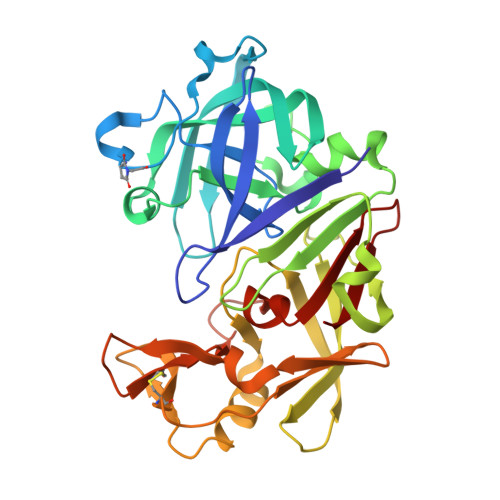
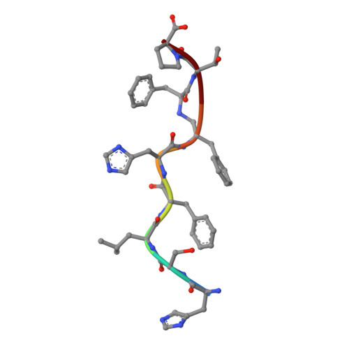
    The analysis reported here describes detailed structural studies of endothiapepsin (the aspartic proteinase from Endothia parasitica), with and without bound inhibitors, and human pepsin 3b. Comparison of multiple crystal structures of members of the aspartic proteinase family has revealed small but significant differences in domain orientation in different crystal forms. In this paper, it is shown that these differences in domain orientation do not necessarily correlate with the presence or absence of bound inhibitors, but appear to stem at least partly from crystal contacts mediated by sulfate ions. However, since the same inherent flexibility of the structure is observed for other enzymes in this family such as human pepsin, the native structure of which is also reported here, the observed domain movements may well have implications for the mechanism of catalysis.


  • Organizational Affiliation
    • Incisive Media, 32-34 Broadwick Street, London W1A 2HG, England.

Macromolecule Content 

  • Total Structure Weight: 35.2 kDa 
  • Atom Count: 2,807 
  • Modeled Residue Count: 337 
  • Deposited Residue Count: 337 
  • Unique protein chains: 2

Macromolecules

Find similar proteins by:|  3D Structure
Entity ID: 1
MoleculeChains  Sequence LengthOrganismDetailsImage
Endothiapepsin329Cryphonectria parasiticaMutation(s): 0 
EC: 3.4.23.22
UniProt
Find proteins for P11838 (Cryphonectria parasitica)
Explore P11838 
Go to UniProtKB:  P11838
Entity Groups
Sequence Clusters30% Identity50% Identity70% Identity90% Identity95% Identity100% Identity
UniProt GroupP11838
Sequence Annotations
Expand
Reference Sequence
Find similar proteins by:  Sequence   |   3D Structure  
Entity ID: 2
MoleculeChains  Sequence LengthOrganismDetailsImage
DB6 peptide8N/AMutation(s): 0 

Biologically Interesting Molecules (External Reference) 

1 Unique

Experimental Data & Validation

Experimental Data

  • Method: X-RAY DIFFRACTION
  • Resolution: 2.00 Å
  • R-Value Free:  0.237 (Depositor), 0.209 (DCC) 
  • R-Value Work:  0.147 (Depositor), 0.132 (DCC) 
  • R-Value Observed: 0.147 (Depositor) 
Space Group: P 1 21 1
Unit Cell:
Length ( Å )Angle ( ˚ )
a = 43.119α = 90
b = 76.02β = 96.99
c = 42.883γ = 90
Software Package:
Software NamePurpose
MAR345data collection
FFTmodel building
SHELXL-97refinement
MOSFLMdata reduction
SCALAdata scaling
FFTphasing

Structure Validation

View Full Validation Report


Entry History 

Deposition Data

Revision History  (Full details and data files)

  • Version 1.0: 2012-04-18
    Type: Initial release
  • Version 1.1: 2012-04-25
    Changes: Database references
  • Version 1.2: 2012-05-09
    Changes: Database references
  • Version 1.3: 2012-12-12
    Changes: Other
  • Version 2.0: 2023-11-15
    Changes: Atomic model, Data collection, Database references, Derived calculations, Refinement description
  • Version 3.0: 2026-03-18
    Changes: Polymer sequence, Structure summary