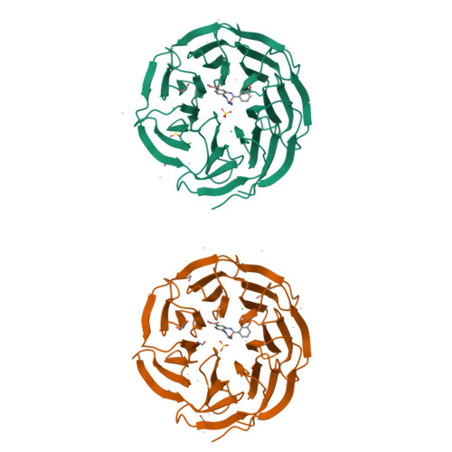
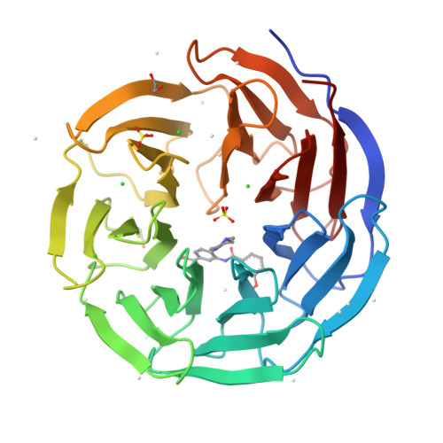
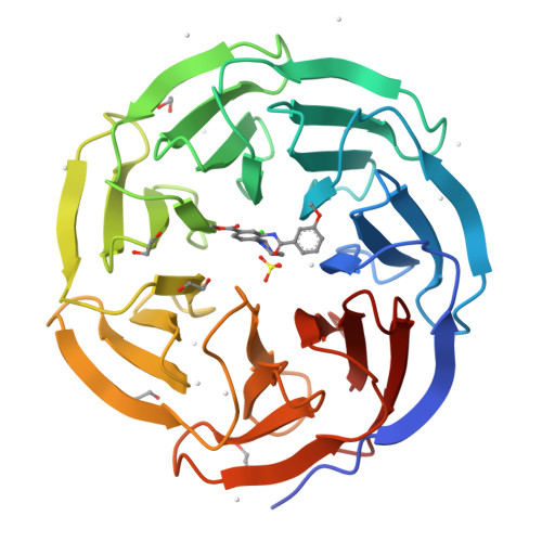
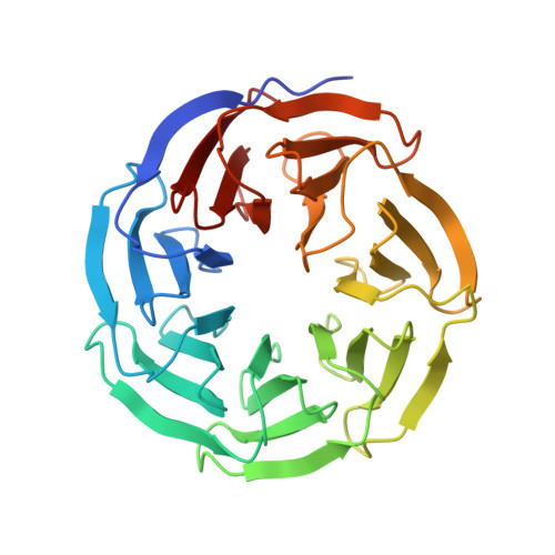
3UR4

Crystal structure of human WD repeat domain 5 with compound


Experimental Data Snapshot

  • Method: X-RAY DIFFRACTION
  • Resolution: 1.80 Å
  • R-Value Free: 0.213 
  • R-Value Work: 0.182 
  • R-Value Observed: 0.182 

Starting Model: experimental
View more details

wwPDB Validation   3D Report Full Report


Ligand Structure Quality Assessment 


This is version 1.3 of the entry. See complete history


Literature

Small-molecule inhibition of MLL activity by disruption of its interaction with WDR5.

Senisterra, G.Wu, H.Allali-Hassani, A.Wasney, G.A.Barsyte-Lovejoy, D.Dombrovski, L.Dong, A.Nguyen, K.T.Smil, D.Bolshan, Y.Hajian, T.He, H.Seitova, A.Chau, I.Li, F.Poda, G.Couture, J.F.Brown, P.J.Al-Awar, R.Schapira, M.Arrowsmith, C.H.Vedadi, M.

(2013) Biochem J 449: 151-159

  • DOI: https://doi.org/10.1042/BJ20121280
  • Primary Citation of Related Structures:  
    3SMR, 3UR4

  • PubMed Abstract: 

    WDR5 (WD40 repeat protein 5) is an essential component of the human trithorax-like family of SET1 [Su(var)3-9 enhancer-of-zeste trithorax 1] methyltransferase complexes that carry out trimethylation of histone 3 Lys4 (H3K4me3), play key roles in development and are abnormally expressed in many cancers. In the present study, we show that the interaction between WDR5 and peptides from the catalytic domain of MLL (mixed-lineage leukaemia protein) (KMT2) can be antagonized with a small molecule. Structural and biophysical analysis show that this antagonist binds in the WDR5 peptide-binding pocket with a Kd of 450 nM and inhibits the catalytic activity of the MLL core complex in vitro. The degree of inhibition was enhanced at lower protein concentrations consistent with a role for WDR5 in directly stabilizing the MLL multiprotein complex. Our data demonstrate inhibition of an important protein-protein interaction and form the basis for further development of inhibitors of WDR5-dependent enzymes implicated in MLL-rearranged leukaemias or other cancers.


  • Organizational Affiliation

    Structural Genomics Consortium, 101 College Street, Toronto, Ontario, Canada.


Macromolecules
Find similar proteins by:  (by identity cutoff)  |  3D Structure
Entity ID: 1
MoleculeChains Sequence LengthOrganismDetailsImage
WD repeat-containing protein 5
A, B
312Homo sapiensMutation(s): 0 
Gene Names: WDR5BIG3
UniProt & NIH Common Fund Data Resources
Find proteins for P61964 (Homo sapiens)
Explore P61964 
Go to UniProtKB:  P61964
PHAROS:  P61964
GTEx:  ENSG00000196363 
Entity Groups  
Sequence Clusters30% Identity50% Identity70% Identity90% Identity95% Identity100% Identity
UniProt GroupP61964
Sequence Annotations
Expand
  • Reference Sequence
Small Molecules
Ligands 5 Unique
IDChains Name / Formula / InChI Key2D Diagram3D Interactions
0BW
Query on 0BW

Download Ideal Coordinates CCD File 
C [auth A],
CA [auth B]
methyl 3-[(3-methoxybenzoyl)amino]-4-(4-methylpiperazin-1-yl)benzoate
C21 H25 N3 O4
ZPLBXOVTSNRBFB-UHFFFAOYSA-N
SO4
Query on SO4

Download Ideal Coordinates CCD File 
J [auth A],
K [auth A],
LA [auth B]
SULFATE ION
O4 S
QAOWNCQODCNURD-UHFFFAOYSA-L
EDO
Query on EDO

Download Ideal Coordinates CCD File 
D [auth A]
DA [auth B]
E [auth A]
EA [auth B]
FA [auth B]
D [auth A],
DA [auth B],
E [auth A],
EA [auth B],
FA [auth B],
GA [auth B],
HA [auth B],
IA [auth B]
1,2-ETHANEDIOL
C2 H6 O2
LYCAIKOWRPUZTN-UHFFFAOYSA-N
CL
Query on CL

Download Ideal Coordinates CCD File 
F [auth A]
G [auth A]
H [auth A]
I [auth A]
JA [auth B]
F [auth A],
G [auth A],
H [auth A],
I [auth A],
JA [auth B],
KA [auth B]
CHLORIDE ION
Cl
VEXZGXHMUGYJMC-UHFFFAOYSA-M
UNX
Query on UNX

Download Ideal Coordinates CCD File 
AA [auth A]
BA [auth A]
L [auth A]
M [auth A]
MA [auth B]
AA [auth A],
BA [auth A],
L [auth A],
M [auth A],
MA [auth B],
N [auth A],
NA [auth B],
O [auth A],
OA [auth B],
P [auth A],
PA [auth B],
Q [auth A],
QA [auth B],
R [auth A],
RA [auth B],
S [auth A],
SA [auth B],
T [auth A],
TA [auth B],
U [auth A],
UA [auth B],
V [auth A],
VA [auth B],
W [auth A],
WA [auth B],
X [auth A],
XA [auth B],
Y [auth A],
Z [auth A]
UNKNOWN ATOM OR ION
X
Binding Affinity Annotations 
IDSourceBinding Affinity
0BW BindingDB:  3UR4 Kd: 3000 (nM) from 1 assay(s)
Experimental Data & Validation

Experimental Data

  • Method: X-RAY DIFFRACTION
  • Resolution: 1.80 Å
  • R-Value Free: 0.213 
  • R-Value Work: 0.182 
  • R-Value Observed: 0.182 
  • Space Group: C 1 2 1
Unit Cell:
Length ( Å )Angle ( ˚ )
a = 134.829α = 90
b = 47.002β = 117.9
c = 112.904γ = 90
Software Package:
Software NamePurpose
SBC-Collectdata collection
PHASERphasing
REFMACrefinement
Cootmodel building
HKL-2000data reduction
HKL-2000data scaling

Structure Validation

View Full Validation Report


Ligand Structure Quality Assessment 


Entry History 

Revision History  (Full details and data files)

  • Version 1.0: 2011-12-14
    Type: Initial release
  • Version 1.1: 2017-11-08
    Changes: Refinement description
  • Version 1.2: 2018-05-16
    Changes: Data collection, Database references
  • Version 1.3: 2023-09-13
    Changes: Data collection, Database references, Derived calculations, Refinement description