3UPC | pdb_00003upc

A general strategy for the generation of human antibody variable domains with increased aggregation resistance


Experimental Data Snapshot

  • Method: X-RAY DIFFRACTION
  • Resolution: 2.80 Å
  • R-Value Free: 
    0.247 (Depositor), 0.243 (DCC) 
  • R-Value Work: 
    0.209 (Depositor), 0.210 (DCC) 
  • R-Value Observed: 
    0.211 (Depositor) 

Starting Model: experimental
View more details

wwPDB Validation 3D Report Full Report

Validation slider image for 3UPC

This is version 1.4 of the entry. See complete history

Literature

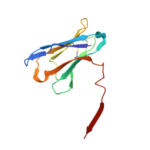
General strategy for the generation of human antibody variable domains with increased aggregation resistance

Dudgeon, K.Rouet, R.Kokmeijer, I.Schofield, P.Stolp, J.Langley, D.Stock, D.Christ, D.

(2012) Proc Natl Acad Sci U S A 109: 10879-10884

  • DOI: https://doi.org/10.1073/pnas.1202866109
  • Primary Citation Related Structures: 
    3UPA, 3UPC

  • PubMed Abstract: 

    The availability of stable human antibody reagents would be of considerable advantage for research, diagnostic, and therapeutic applications. Unfortunately, antibody variable heavy and light domains (V(H) and V(L)) that mediate the interaction with antigen have the propensity to aggregate. Increasing their aggregation resistance in a general manner has proven to be a difficult and persistent problem, due to the high level of sequence diversity observed in human variable domains and the requirement to maintain antigen binding. Here we outline such an approach. By using phage display we identified specific positions that clustered in the antigen binding site (28, 30-33, 35 in V(H) and 24, 49-53, 56 in V(L)). Introduction of aspartate or glutamate at these positions endowed superior biophysical properties (non-aggregating, well-expressed, and heat-refoldable) onto domains derived from common human germline families (V(H)3 and V(κ)1). The effects of the mutations were highly positional and independent of sequence diversity at other positions. Moreover, crystal structures of mutant V(H) and V(L) domains revealed a surprising degree of structural conservation, indicating compatibility with V(H)/V(L) pairing and antigen binding. This allowed the retrofitting of existing binders, as highlighted by the development of robust high affinity antibody fragments derived from the breast cancer therapeutic Herceptin. Our results provide a general strategy for the generation of human antibody variable domains with increased aggregation resistance.


  • Organizational Affiliation
    • Garvan Institute of Medical Research, 384 Victoria Street, Darlinghurst, Sydney, New South Wales 2010, Australia.

Macromolecule Content 

  • Total Structure Weight: 122.43 kDa 
  • Atom Count: 8,217 
  • Modeled Residue Count: 1,130 
  • Deposited Residue Count: 1,130 
  • Unique protein chains: 1

Macromolecules

Find similar proteins by:|  3D Structure
Entity ID: 1
MoleculeChains  Sequence LengthOrganismDetailsImage
heavy chain variable domain
A, B, C, D, E
A, B, C, D, E, F, G, H, I, J
113Homo sapiensMutation(s): 0 
UniProt & NIH Common Fund Data Resources
Find proteins for P01764 (Homo sapiens)
Explore P01764 
Go to UniProtKB:  P01764
PHAROS:  P01764
GTEx:  ENSG00000211949 
Entity Groups
Sequence Clusters30% Identity50% Identity70% Identity90% Identity95% Identity100% Identity
UniProt GroupP01764
Sequence Annotations
Expand
Reference Sequence

Small Molecules

Ligands 4 Unique
IDChains Name / Formula / InChI Key2D Diagram3D Interactions
1PE

Query on 1PE


Download:Ideal Coordinates CCD File
K [auth A],
N [auth D]
PENTAETHYLENE GLYCOL
C10 H22 O6
JLFNLZLINWHATN-UHFFFAOYSA-N
PG4

Query on PG4


Download:Ideal Coordinates CCD File
O [auth G],
P [auth H],
Q [auth I]
TETRAETHYLENE GLYCOL
C8 H18 O5
UWHCKJMYHZGTIT-UHFFFAOYSA-N
PGE

Query on PGE


Download:Ideal Coordinates CCD File
L [auth B]TRIETHYLENE GLYCOL
C6 H14 O4
ZIBGPFATKBEMQZ-UHFFFAOYSA-N
PEG

Query on PEG


Download:Ideal Coordinates CCD File
M [auth C]DI(HYDROXYETHYL)ETHER
C4 H10 O3
MTHSVFCYNBDYFN-UHFFFAOYSA-N

Experimental Data & Validation

Experimental Data

  • Method: X-RAY DIFFRACTION
  • Resolution: 2.80 Å
  • R-Value Free:  0.247 (Depositor), 0.243 (DCC) 
  • R-Value Work:  0.209 (Depositor), 0.210 (DCC) 
  • R-Value Observed: 0.211 (Depositor) 
Space Group: P 21 21 2
Unit Cell:
Length ( Å )Angle ( ˚ )
a = 82.61α = 90
b = 143.12β = 90
c = 145.61γ = 90
Software Package:
Software NamePurpose
MOSFLMdata reduction
SCALAdata scaling
PHASERphasing
REFMACrefinement
PDB_EXTRACTdata extraction
Blu-Icedata collection

Structure Validation

View Full Validation Report


Entry History 

Deposition Data

Revision History  (Full details and data files)

  • Version 1.0: 2012-06-20
    Type: Initial release
  • Version 1.1: 2012-08-15
    Changes: Database references
  • Version 1.2: 2017-11-08
    Changes: Refinement description
  • Version 1.3: 2023-11-01
    Changes: Data collection, Database references, Derived calculations, Refinement description
  • Version 1.4: 2024-11-06
    Changes: Structure summary