3UP3

Nuclear receptor DAF-12 from hookworm Ancylostoma ceylanicum in complex with (25S)-cholestenoic acid


Experimental Data Snapshot

  • Method: X-RAY DIFFRACTION
  • Resolution: 1.25 Å
  • R-Value Free: 
    0.180 (Depositor), 0.190 (DCC) 
  • R-Value Work: 
    0.166 (Depositor), 0.180 (DCC) 
  • R-Value Observed: 
    0.167 (Depositor) 

wwPDB Validation   3D Report Full Report


Ligand Structure Quality Assessment 

Created with Raphaël 2.3.0Worse 01 BetterLigand structure goodness of fit to experimental dataBest fitted XCAClick on this verticalbar to view details

This is version 1.3 of the entry. See complete history


Literature

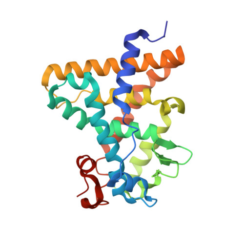
Structural Conservation of Ligand Binding Reveals a Bile Acid-like Signaling Pathway in Nematodes.

Zhi, X.Zhou, X.E.Melcher, K.Motola, D.L.Gelmedin, V.Hawdon, J.Kliewer, S.A.Mangelsdorf, D.J.Xu, H.E.

(2012) J Biological Chem 287: 4894-4903

  • DOI: https://doi.org/10.1074/jbc.M111.315242
  • Primary Citation of Related Structures:  
    3UP0, 3UP3

  • PubMed Abstract: 

    Bile acid-like molecules named dafachronic acids (DAs) control the dauer formation program in Caenorhabditis elegans through the nuclear receptor DAF-12. This mechanism is conserved in parasitic nematodes to regulate their dauer-like infective larval stage, and as such, the DAF-12 ligand binding domain has been identified as an important therapeutic target in human parasitic hookworm species that infect more than 600 million people worldwide. Here, we report two x-ray crystal structures of the hookworm Ancylostoma ceylanicum DAF-12 ligand binding domain in complex with DA and cholestenoic acid (a bile acid-like metabolite), respectively. Structure analysis and functional studies reveal key residues responsible for species-specific ligand responses of DAF-12. Furthermore, DA binds to DAF-12 mechanistically and is structurally similar to bile acids binding to the mammalian bile acid receptor farnesoid X receptor. Activation of DAF-12 by cholestenoic acid and the cholestenoic acid complex structure suggest that bile acid-like signaling pathways have been conserved in nematodes and mammals. Together, these results reveal the molecular mechanism for the interplay between parasite and host, provide a structural framework for DAF-12 as a promising target in treating nematode parasitism, and provide insight into the evolution of gut parasite hormone-signaling pathways.


  • Organizational Affiliation

    Laboratory of Structural Sciences, Van Andel Research Institute, Grand Rapids, Michigan 49503, USA.


Macromolecules
Find similar proteins by:  (by identity cutoff)  |  3D Structure
Entity ID: 1
MoleculeChains Sequence LengthOrganismDetailsImage
aceDAF-12243Ancylostoma ceylanicumMutation(s): 0 
Gene Names: daf-12
UniProt
Find proteins for H0USY8 (Ancylostoma ceylanicum)
Explore H0USY8 
Go to UniProtKB:  H0USY8
Entity Groups  
Sequence Clusters30% Identity50% Identity70% Identity90% Identity95% Identity100% Identity
UniProt GroupH0USY8
Sequence Annotations
Expand
  • Reference Sequence

Find similar proteins by:  Sequence   |   3D Structure  

Entity ID: 2
MoleculeChains Sequence LengthOrganismDetailsImage
Nuclear receptor coactivator 2B [auth P]14Homo sapiensMutation(s): 0 
UniProt & NIH Common Fund Data Resources
Find proteins for Q15596 (Homo sapiens)
Explore Q15596 
Go to UniProtKB:  Q15596
PHAROS:  Q15596
GTEx:  ENSG00000140396 
Entity Groups  
Sequence Clusters30% Identity50% Identity70% Identity90% Identity95% Identity100% Identity
UniProt GroupQ15596
Sequence Annotations
Expand
  • Reference Sequence
Small Molecules
Ligands 2 Unique
IDChains Name / Formula / InChI Key2D Diagram3D Interactions
XCA
Query on XCA

Download Ideal Coordinates CCD File 
C [auth A](8alpha,10alpha,25S)-3-hydroxycholesta-3,5-dien-26-oic acid
C27 H42 O3
NIQOQQMAUFLELB-KMPPVSSLSA-N
EDO
Query on EDO

Download Ideal Coordinates CCD File 
D [auth A]
E [auth A]
F [auth A]
G [auth A]
H [auth A]
1,2-ETHANEDIOL
C2 H6 O2
LYCAIKOWRPUZTN-UHFFFAOYSA-N
Experimental Data & Validation

Experimental Data

  • Method: X-RAY DIFFRACTION
  • Resolution: 1.25 Å
  • R-Value Free:  0.180 (Depositor), 0.190 (DCC) 
  • R-Value Work:  0.166 (Depositor), 0.180 (DCC) 
  • R-Value Observed: 0.167 (Depositor) 
Space Group: P 1 21 1
Unit Cell:
Length ( Å )Angle ( ˚ )
a = 34.545α = 90
b = 85.296β = 106.87
c = 46.325γ = 90
Software Package:
Software NamePurpose
DENZOdata reduction
PHASERphasing
REFMACrefinement
PDB_EXTRACTdata extraction
HKL-2000data collection
SCALEPACKdata scaling

Structure Validation

View Full Validation Report


Ligand Structure Quality Assessment 

Created with Raphaël 2.3.0Worse 01 BetterLigand structure goodness of fit to experimental dataBest fitted XCAClick on this verticalbar to view details

Entry History 

Deposition Data

Revision History  (Full details and data files)

  • Version 1.0: 2011-12-14
    Type: Initial release
  • Version 1.1: 2012-01-11
    Changes: Database references
  • Version 1.2: 2012-02-29
    Changes: Database references
  • Version 1.3: 2024-02-28
    Changes: Data collection, Database references, Derived calculations