3UNC | pdb_00003unc

Crystal Structure of Bovine Milk Xanthine Dehydrogenase to 1.65A Resolution


Experimental Data Snapshot

  • Method: X-RAY DIFFRACTION
  • Resolution: 1.65 Å
  • R-Value Free: 
    0.196 (Depositor), 0.188 (DCC) 
  • R-Value Work: 
    0.177 (Depositor), 0.166 (DCC) 

Starting Model: experimental
View more details

wwPDB Validation 3D Report Full Report

Validation slider image for 3UNC

Ligand Structure Quality Assessment 


This is version 1.1 of the entry. See complete history

Literature

Protein conformational gating of enzymatic activity in xanthine oxidoreductase.

Ishikita, H.Eger, B.T.Okamoto, K.Nishino, T.Pai, E.F.

(2012) J Am Chem Soc 134: 999-1009

  • DOI: https://doi.org/10.1021/ja207173p
  • Primary Citation Related Structures: 
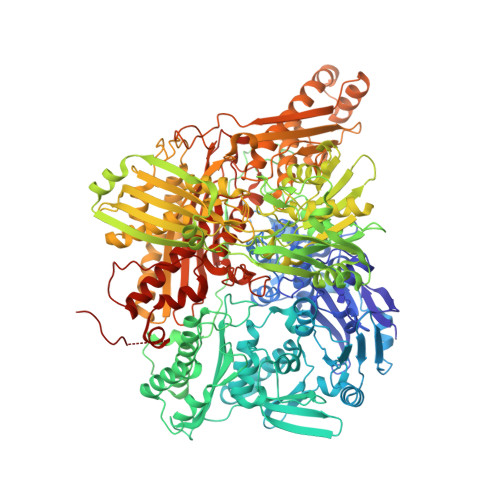
    3AX7, 3AX9, 3UNA, 3UNC, 3UNI

  • PubMed Abstract: 

    In mammals, xanthine oxidoreductase can exist as xanthine dehydrogenase (XDH) and xanthine oxidase (XO). The two enzymes possess common redox active cofactors, which form an electron transfer (ET) pathway terminated by a flavin cofactor. In spite of identical protein primary structures, the redox potential difference between XDH and XO for the flavin semiquinone/hydroquinone pair (E(sq/hq)) is ~170 mV, a striking difference. The former greatly prefers NAD(+) as ultimate substrate for ET from the iron-sulfur cluster FeS-II via flavin while the latter only accepts dioxygen. In XDH (without NAD(+)), however, the redox potential of the electron donor FeS-II is 180 mV higher than that for the acceptor flavin, yielding an energetically uphill ET. On the basis of new 1.65, 2.3, 1.9, and 2.2 Å resolution crystal structures for XDH, XO, the NAD(+)- and NADH-complexed XDH, E(sq/hq) were calculated to better understand how the enzyme activates an ET from FeS-II to flavin. The majority of the E(sq/hq) difference between XDH and XO originates from a conformational change in the loop at positions 423-433 near the flavin binding site, causing the differences in stability of the semiquinone state. There was no large conformational change observed in response to NAD(+) binding at XDH. Instead, the positive charge of the NAD(+) ring, deprotonation of Asp429, and capping of the bulk surface of the flavin by the NAD(+) molecule all contribute to altering E(sq/hq) upon NAD(+) binding to XDH.


  • Organizational Affiliation
    • Career-Path Promotion Unit for Young Life Scientists, Kyoto University, 202 Building E, Graduate School of Medicine, Kyoto 606-8501, Japan. hiro@cp.kyoto-u.ac.jp

Macromolecule Content 

  • Total Structure Weight: 300.01 kDa 
  • Atom Count: 22,475 
  • Modeled Residue Count: 2,575 
  • Deposited Residue Count: 2,664 
  • Unique protein chains: 1

Macromolecules

Find similar proteins by:|  3D Structure
Entity ID: 1
MoleculeChains  Sequence LengthOrganismDetailsImage
Xanthine dehydrogenase/oxidase
A, B
1,332Bos taurusMutation(s): 0 
EC: 1.17.1.4 (PDB Primary Data), 1.17.3.2 (PDB Primary Data)
UniProt
Find proteins for P80457 (Bos taurus)
Explore P80457 
Go to UniProtKB:  P80457
Entity Groups
Sequence Clusters30% Identity50% Identity70% Identity90% Identity95% Identity100% Identity
UniProt GroupP80457
Sequence Annotations
Expand
Reference Sequence

Small Molecules

Ligands 8 Unique
IDChains Name / Formula / InChI Key2D Diagram3D Interactions
FAD

Query on FAD


Download:Ideal Coordinates CCD File
G [auth A],
Z [auth B]
FLAVIN-ADENINE DINUCLEOTIDE
C27 H33 N9 O15 P2
VWWQXMAJTJZDQX-UYBVJOGSSA-N
MTE

Query on MTE


Download:Ideal Coordinates CCD File
E [auth A],
X [auth B]
PHOSPHONIC ACIDMONO-(2-AMINO-5,6-DIMERCAPTO-4-OXO-3,7,8A,9,10,10A-HEXAHYDRO-4H-8-OXA-1,3,9,10-TETRAAZA-ANTHRACEN-7-YLMETHYL)ESTER
C10 H14 N5 O6 P S2
HPEUEJRPDGMIMY-IFQPEPLCSA-N
FES

Query on FES


Download:Ideal Coordinates CCD File
C [auth A],
D [auth A],
V [auth B],
W [auth B]
FE2/S2 (INORGANIC) CLUSTER
Fe2 S2
NIXDOXVAJZFRNF-UHFFFAOYSA-N
MOS

Query on MOS


Download:Ideal Coordinates CCD File
F [auth A],
Y [auth B]
DIOXOTHIOMOLYBDENUM(VI) ION
H Mo O2 S
BDSRWPHSAKXXRG-UHFFFAOYSA-M
SAL

Query on SAL


Download:Ideal Coordinates CCD File
AA [auth B],
H [auth A]
2-HYDROXYBENZOIC ACID
C7 H6 O3
YGSDEFSMJLZEOE-UHFFFAOYSA-N
GOL

Query on GOL


Download:Ideal Coordinates CCD File
BA [auth B]
CA [auth B]
DA [auth B]
EA [auth B]
FA [auth B]
BA [auth B],
CA [auth B],
DA [auth B],
EA [auth B],
FA [auth B],
GA [auth B],
HA [auth B],
I [auth A],
IA [auth B],
J [auth A],
JA [auth B],
K [auth A],
KA [auth B],
L [auth A],
LA [auth B],
M [auth A],
MA [auth B],
N [auth A],
NA [auth B],
O [auth A],
P [auth A],
Q [auth A],
R [auth A],
S [auth A]
GLYCEROL
C3 H8 O3
PEDCQBHIVMGVHV-UHFFFAOYSA-N
CO3

Query on CO3


Download:Ideal Coordinates CCD File
OA [auth B],
T [auth A]
CARBONATE ION
C O3
BVKZGUZCCUSVTD-UHFFFAOYSA-L
CA

Query on CA


Download:Ideal Coordinates CCD File
PA [auth B],
U [auth A]
CALCIUM ION
Ca
BHPQYMZQTOCNFJ-UHFFFAOYSA-N
Binding Affinity Annotations 
IDSourceBinding Affinity
SAL BindingDB:  3UNC Ki: 1.00e+8 (nM) from 1 assay(s)

Experimental Data & Validation

Experimental Data

  • Method: X-RAY DIFFRACTION
  • Resolution: 1.65 Å
  • R-Value Free:  0.196 (Depositor), 0.188 (DCC) 
  • R-Value Work:  0.177 (Depositor), 0.166 (DCC) 
Space Group: C 1 2 1
Unit Cell:
Length ( Å )Angle ( ˚ )
a = 167.573α = 90
b = 124.37β = 91.02
c = 148.177γ = 90
Software Package:
Software NamePurpose
CNSrefinement
HKL-2000data reduction
HKL-2000data scaling
CNSphasing

Structure Validation

View Full Validation Report


Ligand Structure Quality Assessment 


Entry History 

Deposition Data

Revision History  (Full details and data files)

  • Version 1.0: 2012-05-09
    Type: Initial release
  • Version 1.1: 2023-09-13
    Changes: Data collection, Database references, Derived calculations, Refinement description