3UIX | pdb_00003uix

Crystal structure of Pim1 kinase in complex with small molecule inhibitor


Experimental Data Snapshot

  • Method: X-RAY DIFFRACTION
  • Resolution: 2.20 Å
  • R-Value Free: 
    0.201 (Depositor), 0.193 (DCC) 
  • R-Value Work: 
    0.166 (Depositor) 
  • R-Value Observed: 
    0.168 (Depositor) 

Starting Model: experimental
View more details

wwPDB Validation 3D Report Full Report

Validation slider image for 3UIX

Ligand Structure Quality Assessment 


This is version 1.1 of the entry. See complete history

Literature

A novel pim-1 kinase inhibitor targeting residues that bind the substrate Peptide.

Tsuganezawa, K.Watanabe, H.Parker, L.Yuki, H.Taruya, S.Nakagawa, Y.Kamei, D.Mori, M.Ogawa, N.Tomabechi, Y.Handa, N.Honma, T.Yokoyama, S.Kojima, H.Okabe, T.Nagano, T.Tanaka, A.

(2012) J Mol Biology 417: 240-252

  • DOI: https://doi.org/10.1016/j.jmb.2012.01.036
  • Primary Citation Related Structures: 
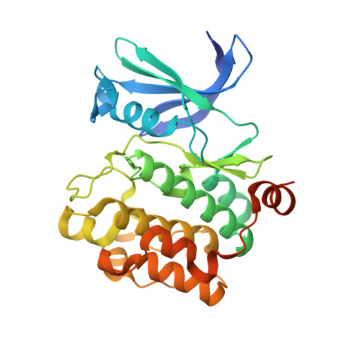
    3UIX

  • PubMed Abstract: 

    A new screening method using fluorescent correlation spectroscopy was developed to select kinase inhibitors that competitively inhibit the binding of a fluorescently labeled substrate peptide. Using the method, among approximately 700 candidate compounds selected by virtual screening, we identified a novel Pim-1 kinase inhibitor targeting its peptide binding residues. X-ray crystal analysis of the complex structure of Pim-1 with the inhibitor indicated that the inhibitor actually binds to the ATP-binding site and also forms direct interactions with residues (Asp128 and Glu171) that bind the substrate peptide. These interactions, which cause small side-chain movements, seem to affect the binding ability of the fluorescently labeled substrate. The compound inhibited Pim-1 kinase in vitro, with an IC(50) value of 150 nM. Treatment of cultured leukemia cells with the compound reduced the amount of p21 and increased the amount of p27, due to Pim-1 inhibition, and then triggered apoptosis after cell-cycle arrest at the G(1)/S phase. This screening method may be widely applicable for the identification of various new Pim-1 kinase inhibitors targeting the residues that bind the substrate peptide.


  • Organizational Affiliation
    • RIKEN Systems and Structural Biology Center, 1-7-22 Suehiro-cho, Tsurumi, Yokohama 230-0045, Japan.

Macromolecule Content 

  • Total Structure Weight: 34.78 kDa 
  • Atom Count: 2,485 
  • Modeled Residue Count: 266 
  • Deposited Residue Count: 298 
  • Unique protein chains: 1

Macromolecules

Find similar proteins by:|  3D Structure
Entity ID: 1
MoleculeChains  Sequence LengthOrganismDetailsImage
Proto-oncogene serine/threonine-protein kinase pim-1298Homo sapiensMutation(s): 0 
Gene Names: PIM1
EC: 2.7.11.1
UniProt & NIH Common Fund Data Resources
Find proteins for P11309 (Homo sapiens)
Explore P11309 
Go to UniProtKB:  P11309
PHAROS:  P11309
GTEx:  ENSG00000137193 
Entity Groups
Sequence Clusters30% Identity50% Identity70% Identity90% Identity95% Identity100% Identity
UniProt GroupP11309
Sequence Annotations
Expand
Reference Sequence

Small Molecules

Ligands 5 Unique
IDChains Name / Formula / InChI Key2D Diagram3D Interactions
Q17

Query on Q17


Download:Ideal Coordinates CCD File
B [auth A]2-{4-[(3-aminopropyl)amino]quinazolin-2-yl}phenol
C17 H18 N4 O
VIQWLKJTZBWAFF-UHFFFAOYSA-N
SO4

Query on SO4


Download:Ideal Coordinates CCD File
E [auth A]SULFATE ION
O4 S
QAOWNCQODCNURD-UHFFFAOYSA-L
GOL

Query on GOL


Download:Ideal Coordinates CCD File
G [auth A]GLYCEROL
C3 H8 O3
PEDCQBHIVMGVHV-UHFFFAOYSA-N
CA

Query on CA


Download:Ideal Coordinates CCD File
F [auth A]CALCIUM ION
Ca
BHPQYMZQTOCNFJ-UHFFFAOYSA-N
CL

Query on CL


Download:Ideal Coordinates CCD File
C [auth A],
D [auth A]
CHLORIDE ION
Cl
VEXZGXHMUGYJMC-UHFFFAOYSA-M

Experimental Data & Validation

Experimental Data

  • Method: X-RAY DIFFRACTION
  • Resolution: 2.20 Å
  • R-Value Free:  0.201 (Depositor), 0.193 (DCC) 
  • R-Value Work:  0.166 (Depositor) 
  • R-Value Observed: 0.168 (Depositor) 
Space Group: P 65
Unit Cell:
Length ( Å )Angle ( ˚ )
a = 97.006α = 90
b = 97.006β = 90
c = 80.898γ = 120
Software Package:
Software NamePurpose
SCALAdata scaling
PHASERphasing
REFMACrefinement
PDB_EXTRACTdata extraction
MOSFLMdata reduction

Structure Validation

View Full Validation Report


Ligand Structure Quality Assessment 


Entry History 

Deposition Data

  • Released Date: 2012-04-11 
  • Deposition Author(s): Parker, L.J.

Revision History  (Full details and data files)

  • Version 1.0: 2012-04-11
    Type: Initial release
  • Version 1.1: 2023-11-01
    Changes: Data collection, Database references, Derived calculations, Refinement description