3UID | pdb_00003uid

Crystal Structure of Protein Ms6760 from Mycobacterium smegmatis


Experimental Data Snapshot

  • Method: X-RAY DIFFRACTION
  • Resolution: 1.57 Å
  • R-Value Free: 
    0.213 (Depositor), 0.208 (DCC) 
  • R-Value Work: 
    0.192 (Depositor), 0.187 (DCC) 
  • R-Value Observed: 
    0.194 (Depositor) 

Starting Model: experimental
View more details

wwPDB Validation 3D Report Full Report

Validation slider image for 3UID

This is version 1.2 of the entry. See complete history

Literature

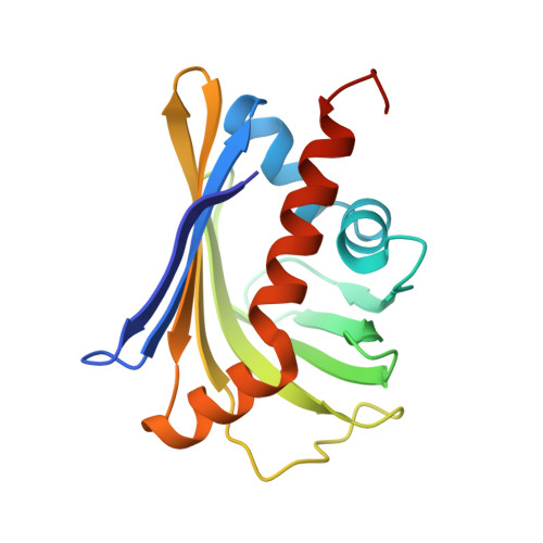
Crystal structure of the toxin Msmeg_6760, the structural homolog of Mycobacterium tuberculosis Rv2035, a novel type II toxin involved in the hypoxic response.

Bajaj, R.A.Arbing, M.A.Shin, A.Cascio, D.Miallau, L.

(2016) Acta Crystallogr F Struct Biol Commun 72: 863-869

  • DOI: https://doi.org/10.1107/S2053230X16017957
  • Primary Citation Related Structures: 
    3UID

  • PubMed Abstract: 

    The structure of Msmeg_6760, a protein of unknown function, has been determined. Biochemical and bioinformatics analyses determined that Msmeg_6760 interacts with a protein encoded in the same operon, Msmeg_6762, and predicted that the operon is a toxin-antitoxin (TA) system. Structural comparison of Msmeg_6760 with proteins of known function suggests that Msmeg_6760 binds a hydrophobic ligand in a buried cavity lined by large hydrophobic residues. Access to this cavity could be controlled by a gate-latch mechanism. The function of the Msmeg_6760 toxin is unknown, but structure-based predictions revealed that Msmeg_6760 and Msmeg_6762 are homologous to Rv2034 and Rv2035, a predicted novel TA system involved in Mycobacterium tuberculosis latency during macrophage infection. The Msmeg_6760 toxin fold has not been previously described for bacterial toxins and its unique structural features suggest that toxin activation is likely to be mediated by a novel mechanism.


  • Organizational Affiliation
    • UCLA-DOE Institute and Departments of Biological Chemistry and Chemistry and Biochemistry, University of California, Los Angeles, CA 90095-1570, USA.

Macromolecule Content 

  • Total Structure Weight: 37.13 kDa 
  • Atom Count: 2,733 
  • Modeled Residue Count: 320 
  • Deposited Residue Count: 336 
  • Unique protein chains: 1

Macromolecules

Find similar proteins by:|  3D Structure
Entity ID: 1
MoleculeChains  Sequence LengthOrganismDetailsImage
Putative uncharacterized protein
A, B
168Mycolicibacterium smegmatis MC2 155Mutation(s): 0 
Gene Names: MSMEG_6760
UniProt
Find proteins for A0R731 (Mycolicibacterium smegmatis (strain ATCC 700084 / mc(2)155))
Explore A0R731 
Go to UniProtKB:  A0R731
Entity Groups
Sequence Clusters30% Identity50% Identity70% Identity90% Identity95% Identity100% Identity
UniProt GroupA0R731
Sequence Annotations
Expand
Reference Sequence

Experimental Data & Validation

Experimental Data

  • Method: X-RAY DIFFRACTION
  • Resolution: 1.57 Å
  • R-Value Free:  0.213 (Depositor), 0.208 (DCC) 
  • R-Value Work:  0.192 (Depositor), 0.187 (DCC) 
  • R-Value Observed: 0.194 (Depositor) 
Space Group: P 21 21 21
Unit Cell:
Length ( Å )Angle ( ˚ )
a = 48.32α = 90
b = 60.21β = 90
c = 100.98γ = 90
Software Package:
Software NamePurpose
XSCALEdata scaling
MOLREPphasing
PHENIXrefinement
PDB_EXTRACTdata extraction
XDSdata reduction

Structure Validation

View Full Validation Report


Entry History 

Deposition Data

Revision History  (Full details and data files)

  • Version 1.0: 2011-11-23
    Type: Initial release
  • Version 1.1: 2016-12-14
    Changes: Database references
  • Version 1.2: 2023-09-13
    Changes: Data collection, Database references, Refinement description