
 Download Mendeley
0.89 A resolution crystal structure of human Parvulin 14 in complex with oxidized DTT
Mueller, J.W., Link, N.M., Matena, A., Hoppstock, L., Rueppel, A., Bayer, P., Blankenfeldt, W.To be published.
Experimental Data Snapshot
Entity ID: 1 | |||||
|---|---|---|---|---|---|
| Molecule | Chains | Sequence Length | Organism | Details | Image |
| Peptidyl-prolyl cis-trans isomerase NIMA-interacting 4 | 101 | Homo sapiens | Mutation(s): 0 Gene Names: PIN4, Q9Y237 EC: 5.2.1.8 |  | |
UniProt & NIH Common Fund Data Resources | |||||
PHAROS: Q9Y237 GTEx: ENSG00000102309 | |||||
Entity Groups | |||||
| Sequence Clusters | 30% Identity50% Identity70% Identity90% Identity95% Identity100% Identity | ||||
| UniProt Group | Q9Y237 | ||||
Sequence AnnotationsExpand | |||||
Reference Sequence | |||||
| Ligands 3 Unique | |||||
|---|---|---|---|---|---|
| ID | Chains | Name / Formula / InChI Key | 2D Diagram | 3D Interactions | |
| D1D Download:Ideal Coordinates CCD File | G [auth A] | (4S,5S)-1,2-DITHIANE-4,5-DIOL C4 H8 O2 S2 YPGMOWHXEQDBBV-QWWZWVQMSA-N | 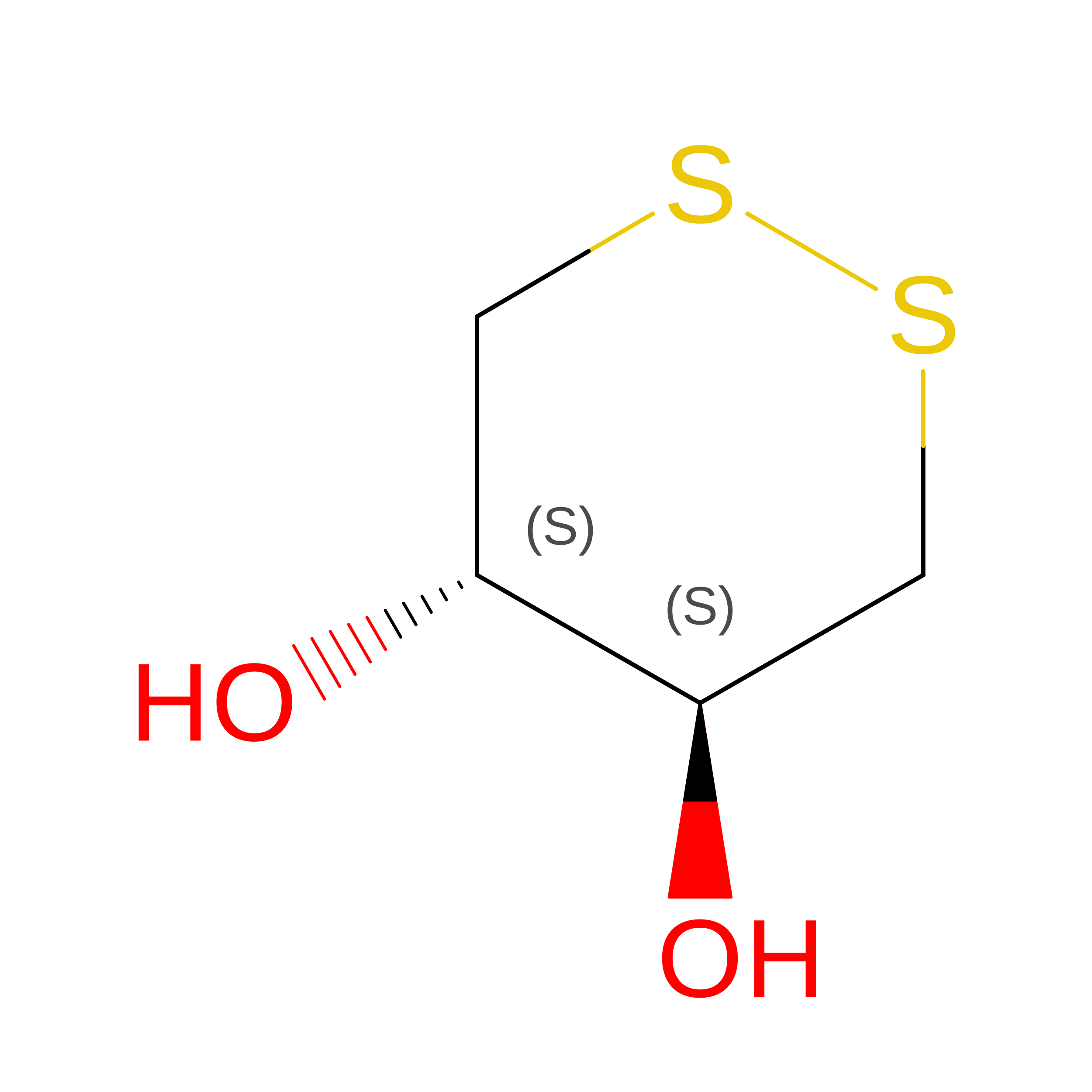 | ||
| SO4 Download:Ideal Coordinates CCD File | B [auth A], C [auth A], D [auth A], E [auth A], F [auth A] | SULFATE ION O4 S QAOWNCQODCNURD-UHFFFAOYSA-L |  | ||
| NA Download:Ideal Coordinates CCD File | H [auth A], I [auth A] | SODIUM ION Na FKNQFGJONOIPTF-UHFFFAOYSA-N |  | ||
| Length ( Å ) | Angle ( ˚ ) |
|---|---|
| a = 34.24 | α = 90 |
| b = 47.02 | β = 90 |
| c = 51.85 | γ = 90 |
| Software Name | Purpose |
|---|---|
| XSCALE | data scaling |
| REFMAC | refinement |
| PDB_EXTRACT | data extraction |
