3UGP | pdb_00003ugp

Crystal structure of RNA-polymerase sigma subunit domain 2 complexed with -10 promoter element ssDNA oligo (TATAAT)


Experimental Data Snapshot

  • Method: X-RAY DIFFRACTION
  • Resolution: 2.70 Å
  • R-Value Free: 
    0.238 (Depositor), 0.231 (DCC) 
  • R-Value Work: 
    0.196 (Depositor), 0.194 (DCC) 
  • R-Value Observed: 
    0.198 (Depositor) 

Starting Model: experimental
View more details

wwPDB Validation 3D Report Full Report

Validation slider image for 3UGP

This is version 1.2 of the entry. See complete history

Literature

Structural basis for promoter-10 element recognition by the bacterial RNA polymerase sigma subunit.

Feklistov, A.Darst, S.A.

(2011) Cell 147: 1257-1269

  • DOI: https://doi.org/10.1016/j.cell.2011.10.041
  • Primary Citation Related Structures: 
    3UGO, 3UGP

  • PubMed Abstract: 

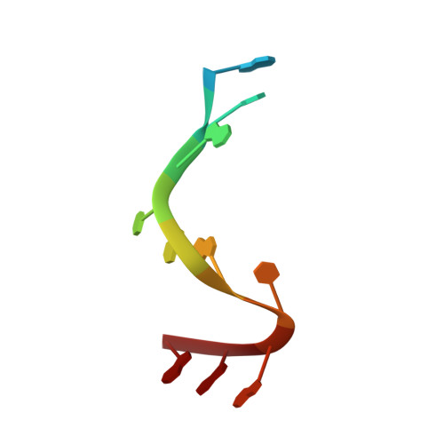
    The key step in bacterial promoter opening is recognition of the -10 promoter element (T(-12)A(-11)T(-10)A(-9)A(-8)T(-7) consensus sequence) by the RNA polymerase σ subunit. We determined crystal structures of σ domain 2 bound to single-stranded DNA bearing-10 element sequences. Extensive interactions occur between the protein and the DNA backbone of every -10 element nucleotide. Base-specific interactions occur primarily with A(-11) and T(-7), which are flipped out of the single-stranded DNA base stack and buried deep in protein pockets. The structures, along with biochemical data, support a model where the recognition of the -10 element sequence drives initial promoter opening as the bases of the nontemplate strand are extruded from the DNA double-helix and captured by σ. These results provide a detailed structural basis for the critical roles of A(-11) and T(-7) in promoter melting and reveal important insights into the initiation of transcription bubble formation.


  • Organizational Affiliation
    • The Rockefeller University, 1230 York Avenue, New York, NY 10065, USA. afeklistov@rockefeller.edu

Macromolecule Content 

  • Total Structure Weight: 35.09 kDa 
  • Atom Count: 1,837 
  • Modeled Residue Count: 194 
  • Deposited Residue Count: 267 
  • Unique protein chains: 1
  • Unique nucleic acid chains: 1

Macromolecules


Find similar proteins by:|  3D Structure
Entity ID: 1
MoleculeChains  Sequence LengthOrganismDetailsImage
RNA polymerase sigma factor245Thermus aquaticusMutation(s): 0 
UniProt
Find proteins for Q9EZJ8 (Thermus aquaticus)
Explore Q9EZJ8 
Go to UniProtKB:  Q9EZJ8
Entity Groups
Sequence Clusters30% Identity50% Identity70% Identity90% Identity95% Identity100% Identity
UniProt GroupQ9EZJ8
Sequence Annotations
Expand
Reference Sequence
Find similar nucleic acids by:  Sequence
Entity ID: 2
MoleculeChains LengthOrganismImage
5'-D(*TP*GP*TP*AP*TP*AP*AP*TP*GP*GP*G)-3'
B, C
11N/A
Sequence Annotations
Expand
Reference Sequence

Experimental Data & Validation

Experimental Data

  • Method: X-RAY DIFFRACTION
  • Resolution: 2.70 Å
  • R-Value Free:  0.238 (Depositor), 0.231 (DCC) 
  • R-Value Work:  0.196 (Depositor), 0.194 (DCC) 
  • R-Value Observed: 0.198 (Depositor) 
Space Group: P 4 2 2
Unit Cell:
Length ( Å )Angle ( ˚ )
a = 86.001α = 90
b = 86.001β = 90
c = 105.862γ = 90
Software Package:
Software NamePurpose
PHENIXmodel building
PHENIXrefinement
HKL-2000data reduction
HKL-2000data scaling
PHENIXphasing

Structure Validation

View Full Validation Report


Entry History 

Deposition Data

Revision History  (Full details and data files)

  • Version 1.0: 2011-12-07
    Type: Initial release
  • Version 1.1: 2012-03-07
    Changes: Database references
  • Version 1.2: 2023-09-13
    Changes: Data collection, Database references, Derived calculations, Refinement description