3UG4 | pdb_00003ug4

Crystal structure of alpha-L-arabinofuranosidase from Thermotoga maritima arabinose complex


Experimental Data Snapshot

  • Method: X-RAY DIFFRACTION
  • Resolution: 2.15 Å
  • R-Value Free: 
    0.207 (Depositor), 0.206 (DCC) 
  • R-Value Work: 
    0.167 (Depositor), 0.167 (DCC) 
  • R-Value Observed: 
    0.169 (Depositor) 

Starting Model: experimental
View more details

wwPDB Validation 3D Report Full Report

Validation slider image for 3UG4

Ligand Structure Quality Assessment 


This is version 2.1 of the entry. See complete history

Literature

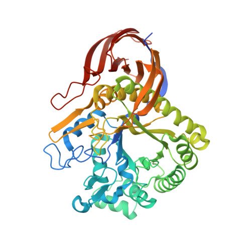
Crystal Structures of Glycoside Hydrolase Family 51 alpha-L-Arabinofuranosidase from Thermotoga maritima

Im, D.-H.Kimura, K.I.Hayasaka, F.Tanaka, T.Noguchi, M.Kobayashi, A.Shoda, S.Miyazaki, K.Wakagi, T.Fushinobu, S.

(2012) Biosci Biotechnol Biochem 76: 423-428

  • DOI: https://doi.org/10.1271/bbb.110902
  • Primary Citation Related Structures: 
    3UG3, 3UG4, 3UG5

  • PubMed Abstract: 

    α-L-Arabinofuranosidase from the hyperthermophilic bacterium Thermotoga maritima (Tm-AFase) is an extremely thermophilic enzyme belonging to glycoside hydrolase family 51. It can catalyze the transglycosylation of a novel glycosyl donor, 4,6-dimethoxy-1,3,5-triazin-2-yl (DMT)-β-D-xylopyranoside. In this study we determined the crystal structures of Tm-AFase in substrate-free and complex forms with arabinose and xylose at 1.8-2.3 Å resolution to determine the architecture of the substrate binding pocket. Subsite -1 of Tm-AFase is similar to that of α-L-arabinofuranosidase from Geobacillus stearothermophilus, but the substrate binding pocket of Tm-AFase is narrower and more hydrophobic. Possible substrate binding modes were investigated by automated docking analysis.


  • Organizational Affiliation
    • Department of Biotechnology, The University of Tokyo, Tokyo, Japan.

Macromolecule Content 

  • Total Structure Weight: 350.43 kDa 
  • Atom Count: 25,486 
  • Modeled Residue Count: 2,893 
  • Deposited Residue Count: 3,024 
  • Unique protein chains: 1

Macromolecules

Find similar proteins by:|  3D Structure
Entity ID: 1
MoleculeChains  Sequence LengthOrganismDetailsImage
Alpha-L-arabinofuranosidase
A, B, C, D, E
A, B, C, D, E, F
504Thermotoga maritimaMutation(s): 1 
Gene Names: TM_0281
EC: 3.2.1.55
UniProt
Find proteins for Q9WYB7 (Thermotoga maritima (strain ATCC 43589 / DSM 3109 / JCM 10099 / NBRC 100826 / MSB8))
Explore Q9WYB7 
Go to UniProtKB:  Q9WYB7
Entity Groups
Sequence Clusters30% Identity50% Identity70% Identity90% Identity95% Identity100% Identity
UniProt GroupQ9WYB7
Sequence Annotations
Expand
Reference Sequence

Small Molecules

Ligands 2 Unique
IDChains Name / Formula / InChI Key2D Diagram3D Interactions
AHR

Query on AHR


Download:Ideal Coordinates CCD File
AA [auth C]
BA [auth C]
CA [auth D]
FA [auth D]
GA [auth D]
AA [auth C],
BA [auth C],
CA [auth D],
FA [auth D],
GA [auth D],
HA [auth D],
I [auth A],
IA [auth D],
J [auth A],
JA [auth D],
K [auth A],
L [auth A],
M [auth A],
MA [auth E],
N [auth A],
NA [auth E],
O [auth A],
OA [auth E],
R [auth B],
RA [auth F],
S [auth B],
SA [auth F],
T [auth B],
TA [auth F],
U [auth B],
UA [auth F],
V [auth B],
VA [auth F],
X [auth C],
Y [auth C],
Z [auth C]
alpha-L-arabinofuranose
C5 H10 O5
HMFHBZSHGGEWLO-QMKXCQHVSA-N
TRS

Query on TRS


Download:Ideal Coordinates CCD File
DA [auth D]
EA [auth D]
G [auth A]
H [auth A]
KA [auth E]
DA [auth D],
EA [auth D],
G [auth A],
H [auth A],
KA [auth E],
LA [auth E],
P [auth B],
PA [auth F],
Q [auth B],
QA [auth F],
W [auth C]
2-AMINO-2-HYDROXYMETHYL-PROPANE-1,3-DIOL
C4 H12 N O3
LENZDBCJOHFCAS-UHFFFAOYSA-O

Experimental Data & Validation

Experimental Data

  • Method: X-RAY DIFFRACTION
  • Resolution: 2.15 Å
  • R-Value Free:  0.207 (Depositor), 0.206 (DCC) 
  • R-Value Work:  0.167 (Depositor), 0.167 (DCC) 
  • R-Value Observed: 0.169 (Depositor) 
Space Group: P 1 21 1
Unit Cell:
Length ( Å )Angle ( ˚ )
a = 100.483α = 90
b = 160.639β = 92.44
c = 154.841γ = 90
Software Package:
Software NamePurpose
HKL-2000data collection
MOLREPphasing
REFMACrefinement
HKL-2000data reduction
HKL-2000data scaling

Structure Validation

View Full Validation Report


Ligand Structure Quality Assessment 


Entry History 

Deposition Data

Revision History  (Full details and data files)

  • Version 1.0: 2012-03-07
    Type: Initial release
  • Version 2.0: 2020-07-29
    Type: Remediation
    Reason: Carbohydrate remediation
    Changes: Atomic model, Data collection, Database references, Derived calculations, Structure summary
  • Version 2.1: 2023-11-01
    Changes: Data collection, Database references, Refinement description, Structure summary