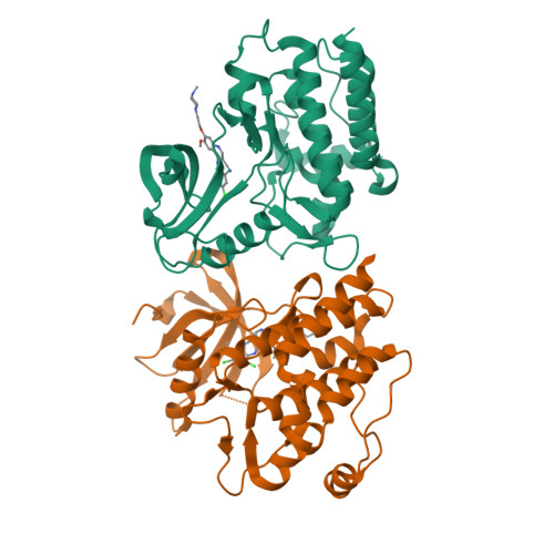
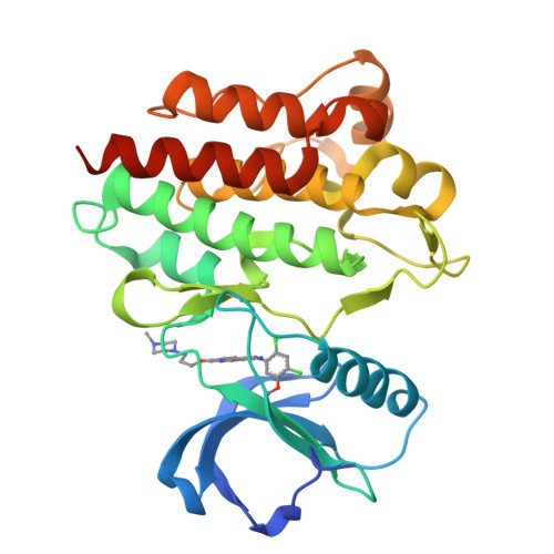
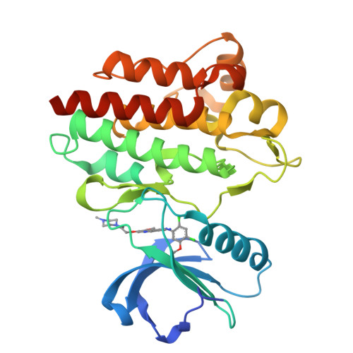
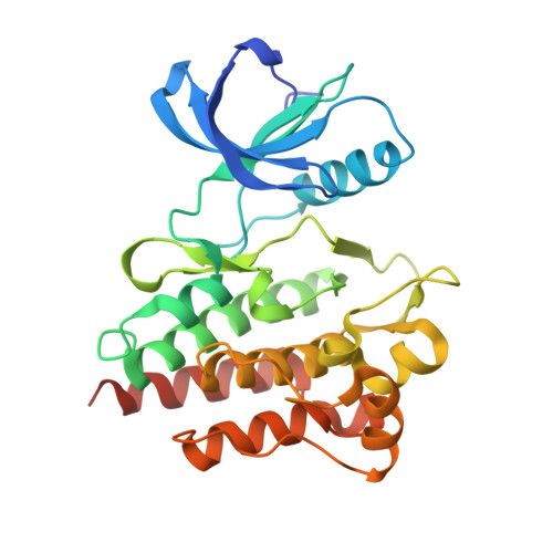
3UE4

Structural and spectroscopic analysis of the kinase inhibitor bosutinib binding to the Abl tyrosine kinase domain


Experimental Data Snapshot

  • Method: X-RAY DIFFRACTION
  • Resolution: 2.42 Å
  • R-Value Free: 0.249 
  • R-Value Work: 0.188 
  • R-Value Observed: 0.192 

wwPDB Validation   3D Report Full Report


Ligand Structure Quality Assessment 


This is version 1.2 of the entry. See complete history


Literature

Structural and spectroscopic analysis of the kinase inhibitor bosutinib and an isomer of bosutinib binding to the abl tyrosine kinase domain.

Levinson, N.M.Boxer, S.G.

(2012) PLoS One 7: e29828-e29828

  • DOI: https://doi.org/10.1371/journal.pone.0029828
  • Primary Citation of Related Structures:  
    3UE4

  • PubMed Abstract: 

    Chronic myeloid leukemia (CML) is caused by the kinase activity of the BCR-Abl fusion protein. The Abl inhibitors imatinib, nilotinib and dasatinib are currently used to treat CML, but resistance to these inhibitors is a significant clinical problem. The kinase inhibitor bosutinib has shown efficacy in clinical trials for imatinib-resistant CML, but its binding mode is unknown. We present the 2.4 Å structure of bosutinib bound to the kinase domain of Abl, which explains the inhibitor's activity against several imatinib-resistant mutants, and reveals that similar inhibitors that lack a nitrile moiety could be effective against the common T315I mutant. We also report that two distinct chemical compounds are currently being sold under the name "bosutinib", and report spectroscopic and structural characterizations of both. We show that the fluorescence properties of these compounds allow inhibitor binding to be measured quantitatively, and that the infrared absorption of the nitrile group reveals a different electrostatic environment in the conserved ATP-binding sites of Abl and Src kinases. Exploiting such differences could lead to inhibitors with improved selectivity.


  • Organizational Affiliation

    Department of Chemistry, Stanford University, Stanford, California, United States of America. nickl@stanford.edu


Macromolecules
Find similar proteins by:  (by identity cutoff)  |  3D Structure
Entity ID: 1
MoleculeChains Sequence LengthOrganismDetailsImage
Tyrosine-protein kinase ABL1
A, B
287Homo sapiensMutation(s): 0 
Gene Names: ABL1ABLJTK7
EC: 2.7.10.2
UniProt & NIH Common Fund Data Resources
Find proteins for P00519 (Homo sapiens)
Explore P00519 
Go to UniProtKB:  P00519
PHAROS:  P00519
GTEx:  ENSG00000097007 
Entity Groups  
Sequence Clusters30% Identity50% Identity70% Identity90% Identity95% Identity100% Identity
UniProt GroupP00519
Sequence Annotations
Expand
  • Reference Sequence
Small Molecules
Ligands 1 Unique
IDChains Name / Formula / InChI Key2D Diagram3D Interactions
DB8
Query on DB8

Download Ideal Coordinates CCD File 
C [auth A],
D [auth B]
4-[(2,4-dichloro-5-methoxyphenyl)amino]-6-methoxy-7-[3-(4-methylpiperazin-1-yl)propoxy]quinoline-3-carbonitrile
C26 H29 Cl2 N5 O3
UBPYILGKFZZVDX-UHFFFAOYSA-N
Binding Affinity Annotations 
IDSourceBinding Affinity
DB8 BindingDB:  3UE4 Kd: min: 0.03, max: 21 (nM) from 15 assay(s)
IC50: min: 0.5, max: 20 (nM) from 7 assay(s)
PDBBind:  3UE4 IC50: 1.1 (nM) from 1 assay(s)
Experimental Data & Validation

Experimental Data

  • Method: X-RAY DIFFRACTION
  • Resolution: 2.42 Å
  • R-Value Free: 0.249 
  • R-Value Work: 0.188 
  • R-Value Observed: 0.192 
  • Space Group: P 2 21 21
Unit Cell:
Length ( Å )Angle ( ˚ )
a = 56.86α = 90
b = 113.76β = 90
c = 127.64γ = 90
Software Package:
Software NamePurpose
PHENIXrefinement
MOSFLMdata reduction
SCALAdata scaling

Structure Validation

View Full Validation Report


Ligand Structure Quality Assessment 


Entry History 

Deposition Data

Revision History  (Full details and data files)

  • Version 1.0: 2012-04-18
    Type: Initial release
  • Version 1.1: 2012-04-25
    Changes: Database references
  • Version 1.2: 2024-02-28
    Changes: Data collection, Database references, Derived calculations