3UD1 | pdb_00003ud1

Crystal structure of ZU5A-ZU5B domains of human erythrocyte ankyrin


Experimental Data Snapshot

  • Method: X-RAY DIFFRACTION
  • Resolution: 2.00 Å
  • R-Value Free: 
    0.206 (Depositor), 0.219 (DCC) 
  • R-Value Work: 
    0.171 (Depositor), 0.187 (DCC) 
  • R-Value Observed: 
    0.173 (Depositor) 

Starting Model: experimental
View more details

wwPDB Validation 3D Report Full Report

Validation slider image for 3UD1

This is version 1.2 of the entry. See complete history

Literature

Structurally Similar but Functionally Diverse ZU5 Domains in Human Erythrocyte Ankyrin.

Yasunaga, M.Ipsaro, J.J.Mondragon, A.

(2012) J Mol Biology 417: 336-350

  • DOI: https://doi.org/10.1016/j.jmb.2012.01.041
  • Primary Citation Related Structures: 
    3UD1, 3UD2

  • PubMed Abstract: 

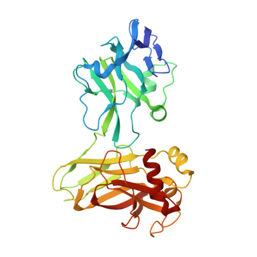
    The metazoan cell membrane is highly organized. Maintaining such organization and preserving membrane integrity under different conditions are accomplished through intracellular tethering to an extensive, flexible protein network. Spectrin, the principal component of this network, is attached to the membrane through the adaptor protein ankyrin, which directly bridges the interaction between β-spectrin and membrane proteins. Ankyrins have a modular structure that includes two tandem ZU5 domains. The first domain, ZU5A, is directly responsible for binding β-spectrin. Here, we present a structure of the tandem ZU5 repeats of human erythrocyte ankyrin. Structural and biophysical experiments show that the second ZU5 domain, ZU5B, does not participate in spectrin binding. ZU5B is structurally similar to the ZU5 domain found in the netrin receptor UNC5b supramodule, suggesting that it could interact with other domains in ankyrin. Comparison of several ZU5 domains demonstrates that the ZU5 domain represents a compact and versatile protein interaction module.


  • Organizational Affiliation
    • Department of Molecular Biosciences, Northwestern University, Evanston, IL 60208, USA.

Macromolecule Content 

  • Total Structure Weight: 108.81 kDa 
  • Atom Count: 8,547 
  • Modeled Residue Count: 966 
  • Deposited Residue Count: 978 
  • Unique protein chains: 1

Macromolecules

Find similar proteins by:|  3D Structure
Entity ID: 1
MoleculeChains  Sequence LengthOrganismDetailsImage
Ankyrin-1
A, B, C
326Homo sapiensMutation(s): 1 
Gene Names: ANK1ANK
UniProt & NIH Common Fund Data Resources
Find proteins for P16157 (Homo sapiens)
Explore P16157 
Go to UniProtKB:  P16157
PHAROS:  P16157
GTEx:  ENSG00000029534 
Entity Groups
Sequence Clusters30% Identity50% Identity70% Identity90% Identity95% Identity100% Identity
UniProt GroupP16157
Sequence Annotations
Expand
Reference Sequence

Small Molecules

Ligands 1 Unique
IDChains Name / Formula / InChI Key2D Diagram3D Interactions
EOH

Query on EOH


Download:Ideal Coordinates CCD File
D [auth A]
E [auth A]
F [auth A]
G [auth A]
H [auth A]
D [auth A],
E [auth A],
F [auth A],
G [auth A],
H [auth A],
I [auth B],
J [auth B],
K [auth B],
L [auth B],
M [auth B],
N [auth C],
O [auth C]
ETHANOL
C2 H6 O
LFQSCWFLJHTTHZ-UHFFFAOYSA-N

Experimental Data & Validation

Experimental Data

  • Method: X-RAY DIFFRACTION
  • Resolution: 2.00 Å
  • R-Value Free:  0.206 (Depositor), 0.219 (DCC) 
  • R-Value Work:  0.171 (Depositor), 0.187 (DCC) 
  • R-Value Observed: 0.173 (Depositor) 
Space Group: C 1 2 1
Unit Cell:
Length ( Å )Angle ( ˚ )
a = 160.123α = 90
b = 40.491β = 112.68
c = 178.104γ = 90
Software Package:
Software NamePurpose
MAR345dtbdata collection
SHARPphasing
BUSTERrefinement
XDSdata reduction
SCALAdata scaling

Structure Validation

View Full Validation Report


Entry History 

Deposition Data

Revision History  (Full details and data files)

  • Version 1.0: 2012-02-22
    Type: Initial release
  • Version 1.1: 2012-03-28
    Changes: Database references
  • Version 1.2: 2023-09-13
    Changes: Data collection, Database references, Derived calculations, Refinement description