3UC5

Phosphopantetheine adenylyltransferase from Mycobacterium tuberculosis complexed with ATP


Experimental Data Snapshot

  • Method: X-RAY DIFFRACTION
  • Resolution: 1.70 Å
  • R-Value Free: 0.222 
  • R-Value Work: 0.177 
  • R-Value Observed: 0.179 

Starting Model: experimental
View more details

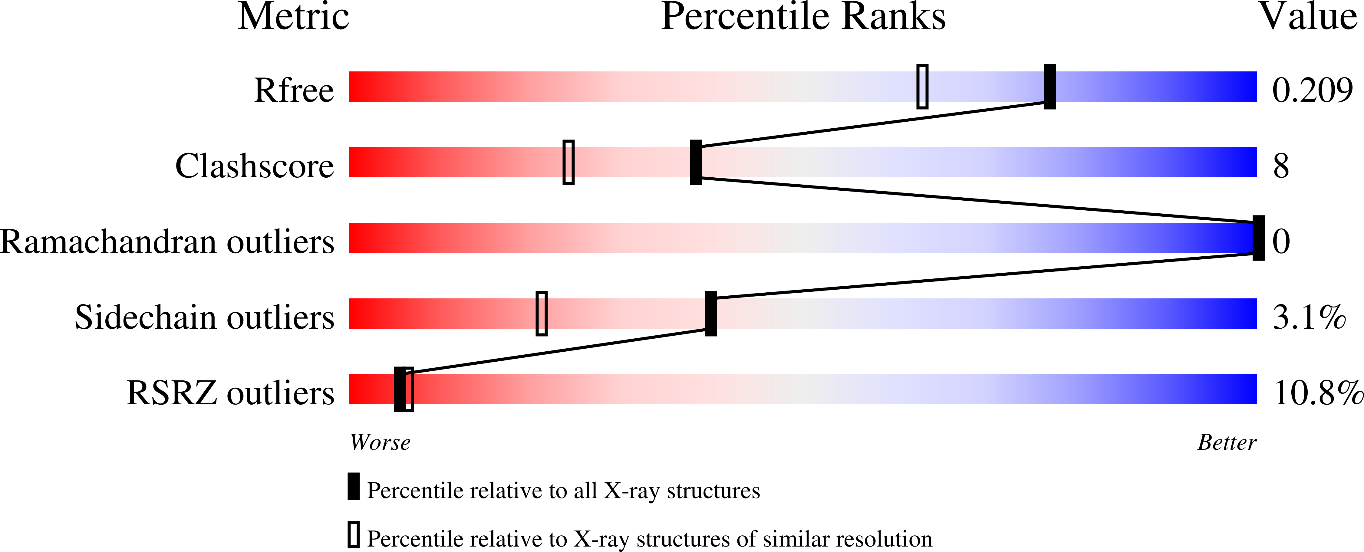
wwPDB Validation   3D Report Full Report


Ligand Structure Quality Assessment 


This is version 1.3 of the entry. See complete history


Literature

X-ray study of the conformational changes in the molecule of phosphopantetheine adenylyltransferase from Mycobacterium tuberculosis during the catalyzed reaction.

Timofeev, V.Smirnova, E.Chupova, L.Esipov, R.Kuranova, I.

(2012) Acta Crystallogr D Biol Crystallogr 68: 1660-1670

  • DOI: https://doi.org/10.1107/S0907444912040206
  • Primary Citation of Related Structures:  
    3UC5, 4E1A

  • PubMed Abstract: 

    Structures of recombinant phosphopantetheine adenylyltransferase (PPAT) from Mycobacterium tuberculosis (PPATMt) in the apo form and in complex with the substrate ATP were determined at 1.62 and 1.70 Å resolution, respectively, using crystals grown in microgravity by the counter-diffusion method. The ATP molecule of the PPATMt-ATP complex was located with full occupancy in the active-site cavity. Comparison of the solved structures with previously determined structures of PPATMt complexed with the reaction product dephosphocoenzyme A (dPCoA) and the feedback inhibitor coenzyme A (CoA) was performed using superposition on C(α) atoms. The peculiarities of the arrangement of the ligands in the active-site cavity of PPATMt are described. The conformational states of the PPAT molecule in the consequent steps of the catalyzed reaction in the apo enzyme and the enzyme-substrate and enzyme-product complexes are characterized. It is shown that the binding of ATP and dPCoA induces the rearrangement of a short part of the polypeptide chain restricting the active-site cavity in the subunits of the hexameric enzyme molecule. The changes in the quaternary structure caused by this rearrangement are accompanied by a variation of the size of the inner water-filled channel which crosses the PPAT molecule along the threefold axis of the hexamer. The molecular mechanism of the observed changes is described.


  • Organizational Affiliation

    Laboratory of X-ray Analysis Methods and Synchrotron Radiation, Shubnikov Institute of Crystallography, Russian Academy of Sciences, Leninsky Prospect 59, Moscow, Russian Federation. tostars@mail.ru


Macromolecules
Find similar proteins by:  (by identity cutoff)  |  3D Structure
Entity ID: 1
MoleculeChains Sequence LengthOrganismDetailsImage
Phosphopantetheine adenylyltransferase157Mycobacterium tuberculosisMutation(s): 0 
Gene Names: coaDkdtBRv2965cMT3043MTCY349.22u0002e
EC: 2.7.7.3
UniProt
Find proteins for P9WPA5 (Mycobacterium tuberculosis (strain ATCC 25618 / H37Rv))
Explore P9WPA5 
Go to UniProtKB:  P9WPA5
Entity Groups  
Sequence Clusters30% Identity50% Identity70% Identity90% Identity95% Identity100% Identity
UniProt GroupP9WPA5
Sequence Annotations
Expand
  • Reference Sequence
Small Molecules
Ligands 1 Unique
IDChains Name / Formula / InChI Key2D Diagram3D Interactions
ATP
Query on ATP

Download Ideal Coordinates CCD File 
B [auth A]ADENOSINE-5'-TRIPHOSPHATE
C10 H16 N5 O13 P3
ZKHQWZAMYRWXGA-KQYNXXCUSA-N
Experimental Data & Validation

Experimental Data

  • Method: X-RAY DIFFRACTION
  • Resolution: 1.70 Å
  • R-Value Free: 0.222 
  • R-Value Work: 0.177 
  • R-Value Observed: 0.179 
  • Space Group: H 3 2
Unit Cell:
Length ( Å )Angle ( ˚ )
a = 99.909α = 90
b = 99.909β = 90
c = 114.419γ = 120
Software Package:
Software NamePurpose
HKL-2000data collection
PHASERphasing
PHENIXrefinement
HKL-2000data reduction
HKL-2000data scaling

Structure Validation

View Full Validation Report


Ligand Structure Quality Assessment 


Entry History 

Deposition Data

Revision History  (Full details and data files)

  • Version 1.0: 2012-11-07
    Type: Initial release
  • Version 1.1: 2012-11-28
    Changes: Database references
  • Version 1.2: 2023-09-13
    Changes: Data collection, Database references, Derived calculations, Refinement description
  • Version 1.3: 2024-11-27
    Changes: Structure summary