3UAG | pdb_00003uag

UDP-N-ACETYLMURAMOYL-L-ALANINE:D-GLUTAMATE LIGASE


Experimental Data Snapshot

  • Method: X-RAY DIFFRACTION
  • Resolution: 1.77 Å
  • R-Value Free: 
    0.222 (Depositor) 
  • R-Value Work: 
    0.178 (Depositor), 0.174 (DCC) 

wwPDB Validation 3D Report Full Report

Validation slider image for 3UAG

Ligand Structure Quality Assessment 


This is version 1.4 of the entry. See complete history

Literature

Determination of the MurD mechanism through crystallographic analysis of enzyme complexes.

Bertrand, J.A.Auger, G.Martin, L.Fanchon, E.Blanot, D.Le Beller, D.van Heijenoort, J.Dideberg, O.

(1999) J Mol Biology 289: 579-590

  • DOI: https://doi.org/10.1006/jmbi.1999.2800
  • Primary Citation Related Structures: 
    2UAG, 3UAG, 4UAG

  • PubMed Abstract: 

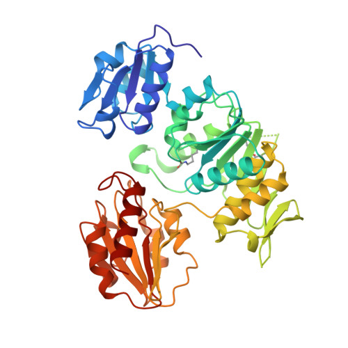
    UDP -N- acetylmuramoyl- L -alanine: D -glutamate (MurD) ligase catalyses the addition of d -glutamate to the nucleotide precursor UDP -N- acetylmuramoyl- L -alanine (UMA). The crystal structures of three complexes of Escherichia coli MurD with a variety of substrates and products have been determined to high resolution. These include (1) the quaternary complex of MurD, the substrate UMA, the product ADP, and Mg2+, (2) the quaternary complex of MurD, the substrate UMA, the product ADP, and Mn2+, and (3) the binary complex of MurD with the product UDP - N- acetylmuramoyl- L -alanine- D -glutamate (UMAG). The reaction mechanism supported by these structures proceeds by the phosphorylation of the C-terminal carboxylate group of UMA by the gamma-phosphate group of ATP to form an acyl-phosphate intermediate, followed by the nucleophilic attack by the amino group of D-glutamate to produce UMAG. A key feature in the reaction intermediate is the presence of two magnesium ions bridging negatively charged groups.


  • Organizational Affiliation
    • Laboratoire de Cristallographie Macromoléculaire, Institut de Biologie Structurale Jean-Pierre Ebel (CEA-CNRS), 41 rue Jules Horowitz, Grenoble Cedex 1, F-38027, France.

Macromolecule Content 

  • Total Structure Weight: 48.4 kDa 
  • Atom Count: 3,690 
  • Modeled Residue Count: 430 
  • Deposited Residue Count: 437 
  • Unique protein chains: 1

Macromolecules

Find similar proteins by:|  3D Structure
Entity ID: 1
MoleculeChains  Sequence LengthOrganismDetailsImage
PROTEIN (UDP-N-ACETYLMURAMOYL-L-ALANINE:D-GLUTAMATE LIGASE)437Escherichia coliMutation(s): 0 
Gene Names: MURD GENE
EC: 6.3.2.9
UniProt
Find proteins for P14900 (Escherichia coli (strain K12))
Explore P14900 
Go to UniProtKB:  P14900
Entity Groups
Sequence Clusters30% Identity50% Identity70% Identity90% Identity95% Identity100% Identity
UniProt GroupP14900
Sequence Annotations
Expand
Reference Sequence

Small Molecules

Modified Residues  1 Unique
IDChains TypeFormula2D DiagramParent
KCX
Query on KCX
A
L-PEPTIDE LINKINGC7 H14 N2 O4LYS

Experimental Data & Validation

Experimental Data

  • Method: X-RAY DIFFRACTION
  • Resolution: 1.77 Å
  • R-Value Free:  0.222 (Depositor) 
  • R-Value Work:  0.178 (Depositor), 0.174 (DCC) 
Space Group: P 41
Unit Cell:
Length ( Å )Angle ( ˚ )
a = 65.24α = 90
b = 65.24β = 90
c = 134.41γ = 90
Software Package:
Software NamePurpose
DENZOdata reduction
SCALEPACKdata scaling
X-PLORrefinement

Structure Validation

View Full Validation Report


Ligand Structure Quality Assessment 


Entry History 

Deposition Data

Revision History  (Full details and data files)

  • Version 1.0: 2000-02-25
    Type: Initial release
  • Version 1.1: 2007-10-16
    Changes: Version format compliance
  • Version 1.2: 2011-07-13
    Changes: Version format compliance
  • Version 1.3: 2019-11-06
    Changes: Data collection, Database references, Derived calculations
  • Version 1.4: 2023-12-27
    Changes: Data collection, Database references, Derived calculations